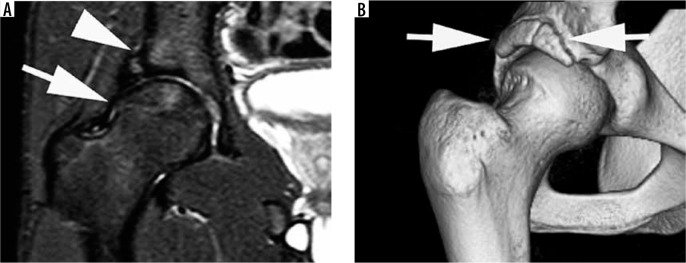
Figure 22.
Mixed cam and pincer femoroacetabular impingement with acetabular rim fracture. A) Coronal proton density fat-saturated magnetic resonance image demonstrating a chronic acetabular rim fracture (short arrow) with over-coverage of the femoral head and cam morphology (long arrow). There is minor subarticular stress change with mild patchy bone marrow oedema at the periphery of the acetabulum and at the proximal aspect of the femoral head. B) 3D computed tomography reformat demonstrating the chronic acetabular rim fracture configuration (arrows)

