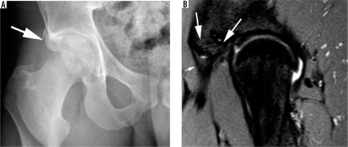
Figure 24.
Subspine impingement. A) Anteroposterior radiograph of the hip demonstrating a type 3 anterior inferior iliac spine (AIIS) configuration ex- tending below the level of the acetabular rim following an old avulsion injury (arrow). B) Sagittal proton density fat-saturated magnetic resonance image demonstrating a malunited AIIS (large arrows). The small arrow indicates the rectus femoris tendon

