
Figure 25.
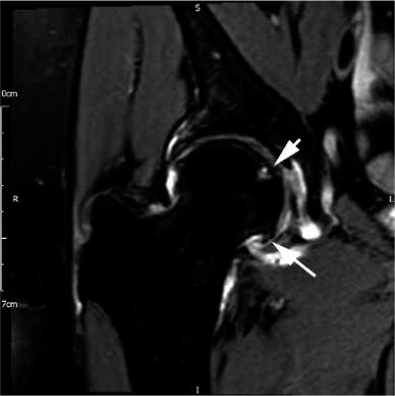
Hip osteoarthritis. Coronal proton density fat-saturated magnetic resonance image demonstrating substantial articular cartilage loss, a joint effusion, and prominent marginal osteophytes (long arrow) with early subchondral cyst-like formation in the femoral head (short arrow)