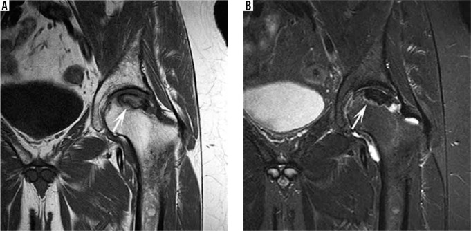
Figure 8.
Osteonecrosis of the femoral head. A) Coronal T1W magnetic resonance (MR) image demonstrating a low/intermediate signal geographic lesion in the subchondral region of the proximal femoral head surrounded by a low signal intensity rim (long arrow). B) Coronal short-tau inversion recovery MR image demonstrating a geographic lesion of low signal intensity in the proximal aspect of the femoral head with an inner line of high signal on the necrotic side repre- senting reparative granulation tissue of the reactive interface and an outer line of low signal representing sclerosis, described as the “double line” sign (long arrow)

