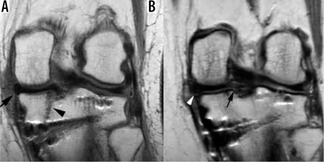
Figure 15.

Meniscal transplant. Coronal proton density-weighted non-fat- saturated contrast enhanced magnetic resonance (MR) images in a 39-year-old female following medial meniscal transplant. A) Postoperative MR image taken 3 months after meniscal transplant shows the meniscal transplant (arrow) and the posterior tibial tunnel (arrowhead). B) MR image obtained two years later, after the patient started experiencing worsening medial knee pain, shows new tearing of the posterior root ligament (arrow) and heterogeneous undersurface signal along the periphery of the body/posterior horn junction (arrowhead), which was found to represent a developing peripheral meniscal tear
