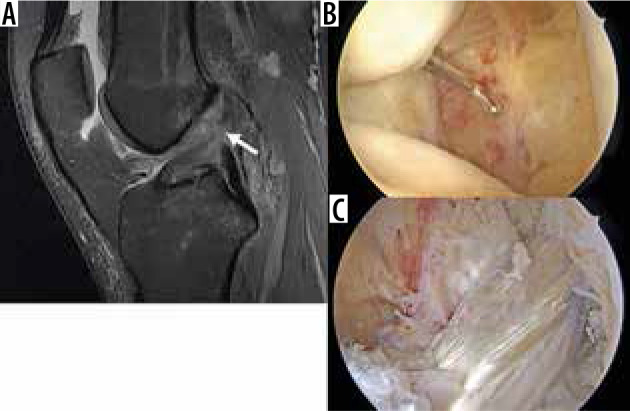
Figure 18.

Full thickness anterior cruciate ligament (ACL) tear and recon- struction. A) Sagittal proton density-weighted fat-saturated magnetic resonance image in a 23-year-old female demonstrates complete discon- tinuity of the proximal to mid ACL (arrow). B) Arthroscopic image confirms complete ACL tear, with ACL stumps visible. C) Subsequent arthroscopic image following ligament graft reconstruction shows an intact hamstring autograft reconstruction
