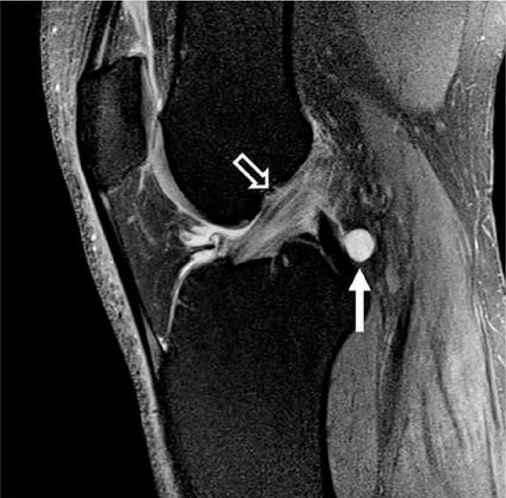
Figure 19.

Mucoid degeneration. Sagittal proton density-weighted fat-satu- rated magnetic resonance image in a 43-year-old male with increased signal within the anterior cruciate ligament (open arrow) with intact fibres repre- sents mucoid degeneration. A cruciate ligament ganglion is incidentally noted posteriorly at the posterior cruciate ligament insertion (closed arrow)
