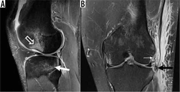
Figure 20.

Secondary signs of anterior cruciate ligament (ACL) tear. A) Sagittal proton density-weighted (PDW) fat-saturated (FS) magnetic resonance (MR) image in a 25-year-old male shows the characteristic osseous contusion pat- tern in ACL injuries from a pivot-shift mechanism, with contusion/impaction along the terminal sulcus of the lateral femoral condyle (open arrow) and the posterior tibial plateau (closed arrow). B) Sagittal PDW FS MR image from a different patient with an ACL injury shows a Segond fracture (black arrow) and injury to the underlying anterolateral ligament (thin, white arrow)
