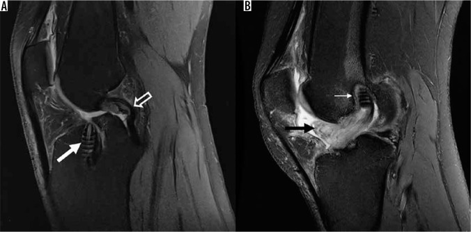
Figure 22.
Anterior cruciate ligament (ACL) reconstruction complications. A) Sagittal proton density-weighted (PDW) fat-saturated (FS) magnetic resonance (MR) image in a 51-year-old male with completely absent ACL graft reconstruction representing tear of ACL reconstruction. There is an abnormally vertically oriented tibial tunnel interference screw present (closed arrow). Note the buckled appearance of the posterior cruciate ligament (open arrow). B) Sagittal PDW FS MR image in an 18-year-old female who is 3 years status post ACL reconstruction, with arthrofibrosis at the anterior aspect of the intercondylar notch, called a “cyclops lesion” (black arrow). A femoral interference screw is partially visible, and there is a thin halo of intermediate signal surrounding it (small arrow), which may represent granulation reaction that can form adjacent to intraosseous device constructed from bioabsorbable materials

