Figure 26.

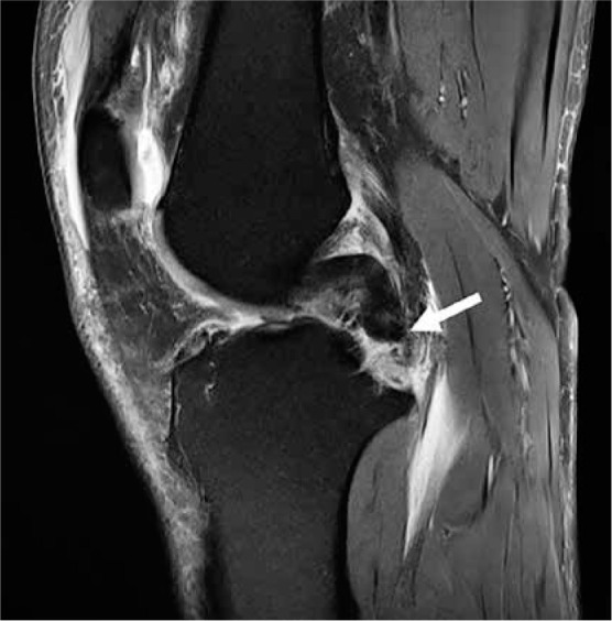
Posterior cruciate ligament (PCL): osseous avulsion. Sagittal proton density-weighted fat-saturated magnetic resonance image in a 54-year-old male shows a complete avulsion of the PCL from its tibial insertion (arrow)

Posterior cruciate ligament (PCL): osseous avulsion. Sagittal proton density-weighted fat-saturated magnetic resonance image in a 54-year-old male shows a complete avulsion of the PCL from its tibial insertion (arrow)