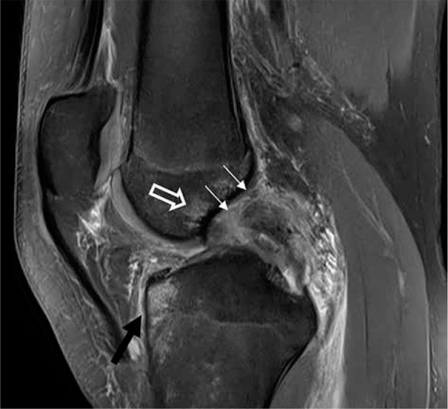
Figure 27.

Posterior cruciate ligament (PCL): complete tear and secondary signs of injury. Sagittal proton density-weighted fat-saturated magnetic resonance image in a 44-year-old male with characteristic osseous contusions in the an- terior proximal tibia (black arrow) and distal femur (open arrow), which occur with PCL injuries. The PCL is thickened, with increased signal and indistinct fibres, and a complete proximal tear from its femoral origin (thin, white arrows)
