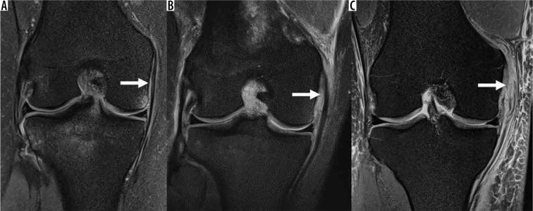
Figure 28.
Medial collateral ligament (MCL) injuries. Coronal proton density-weighted fat-saturated magnetic resonance images in a: A) 20-year-old female with oedema surrounding the MCL without disruption of the ligament fibres (arrow), consistent with a low-grade injury/grade I MCL sprain, B) 21-year-old female with oedema and thickening of the MCL fibres (arrow), consistent with partial-thickness tear/grade II MCL sprain, C) 45-year-old male with oedema and discontinuity of the proximal MCL fibres (arrow), consistent with a full-thickness tear/grade III MCL sprain

