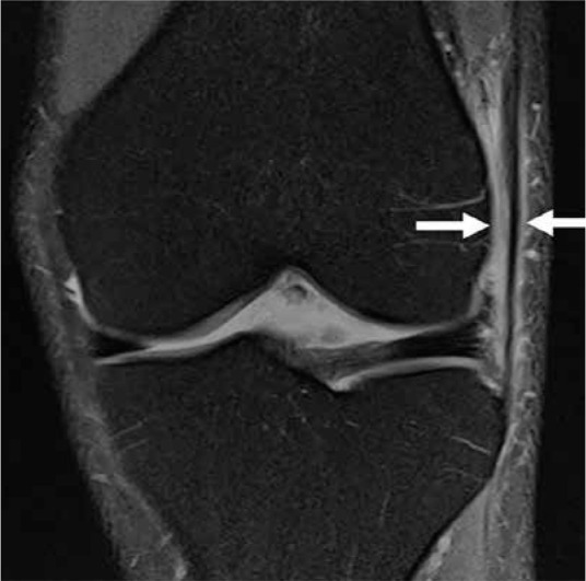
Figure 29.

Iliotibial band syndrome. Coronal proton density-weighted fat-saturated magnetic resonance image of a 34-year-old male with ilio- tibial band friction syndrome, with fluid and oedema adjacent to the lateral femoral condyle and surrounding the iliotibial band (arrows)
