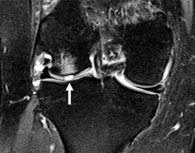
Figure 34.

Cartilage defect. Coronal proton density-weighted fat-saturat- ed magnetic resonance image in a 36-year-old male with a full thickness (grade 4) lateral femoral condyle cartilaginous defect (arrow) and adjacent subchondral bone marrow oedema-like signal
