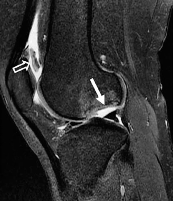
Figure 35.

Osteochondritis dissecans. Sagittal proton density-weighted fat-saturated magnetic resonance image in a 23-year-old female with a large osteochondral defect of the lateral femoral condyle (closed arrow) with adjacent marrow oedema-like signal abnormality. The osteochondral fragment is displaced into the suprapatellar recess (open arrow)
