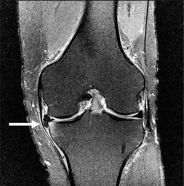
Figure 8.

Horizontal meniscal tear with displaced fragment. Coronal proton densi- ty-weighted fat-saturated magnetic resonance image in a 53-year-old male with tear of the medial meniscus and displaced meniscal fragment/flap (arrow) locat- ed between the medial collateral ligament and the medial tibial plateau. There is subchondral bone marrow oedema-like signal in the adjacent tibia
