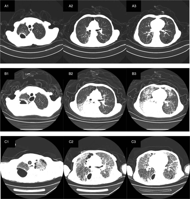
Figure 1.

CT examination of 64 years old man diagnosed with severe COVID-19 pneumonia. (A 1–3) CT showed that there was no beak like air containing cavity in the anastomotic stoma, no communication with the right pleura, and a small amount of air accumulation and effusion in the right chest. Bilateral lungs scattered inflammation with interstitial lesions. Chronic bronchitis, emphysema. (B 1–3) CT showed that the anastomotic stoma seemed to be connected with the right pleural cavity, encapsulated pleural effusion and pneumatosis were more than before, and there were double patchy fuzzy shadows in both lungs. (C 1–3) After 30 days, CT showed that the anastomotic stoma seemed not to be connected with the right pleural cavity, encapsulated pleural effusion and pneumatosis were less than before, multiple patchy fuzzy shadows in double lungs were more absorbed than before, and the bronchial vascular bundles of both lungs were increased, disordered and interstitial thickening.
