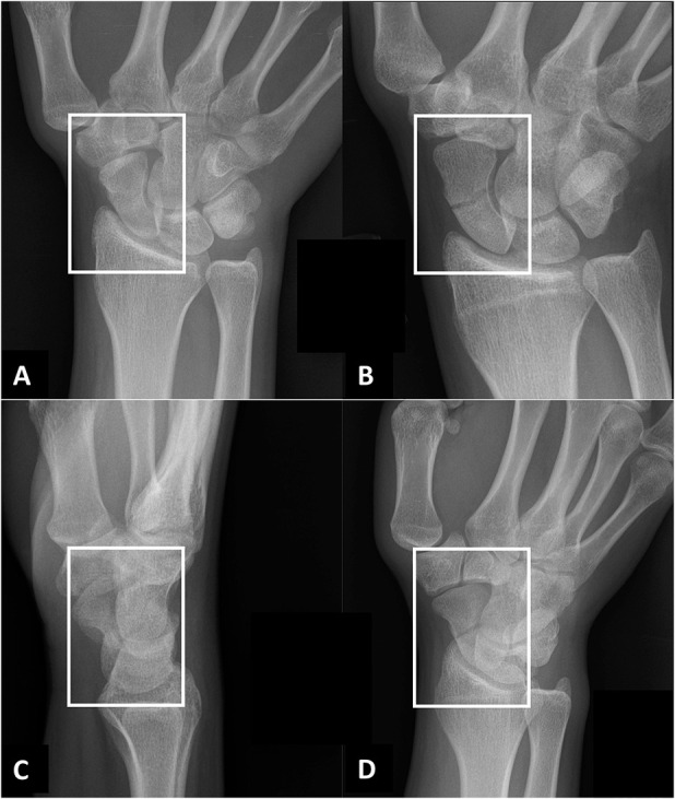
Fig. 1 A-D.

A radiographic scaphoid fracture series for patients with a clinical suspicion for scaphoid fracture at our hospital. The following four projections were fed into the deep learning framework: (A) posterior-anterior ulnar deviation; (B) uptilt (that is, an elongated view with tube angle adjusted over 30°); (C) lateral; and (D) 45° oblique projections. The white boxes illustrate the cropped and resized radiographs (350 x 300 pixels) that are fed into the deep learning framework (VGG 16).
