
Figure 3.
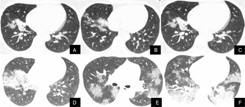
The CT images of a 33-year-old woman with new coronavirus pneumonia shows the progression of the disease. A–B. The first CT scan showed flaky ground glass shadows in the lower lobes of both lungs. C–D. Three days later, the lesions were enlarged, accompanied by consolidation and thickening of interlobular septa. E–F. CT images re-examination 8 days later showed the fusion of large ground glass shadow, consolidation shadow, and interlobar pleural thickening. CT = computed tomography.