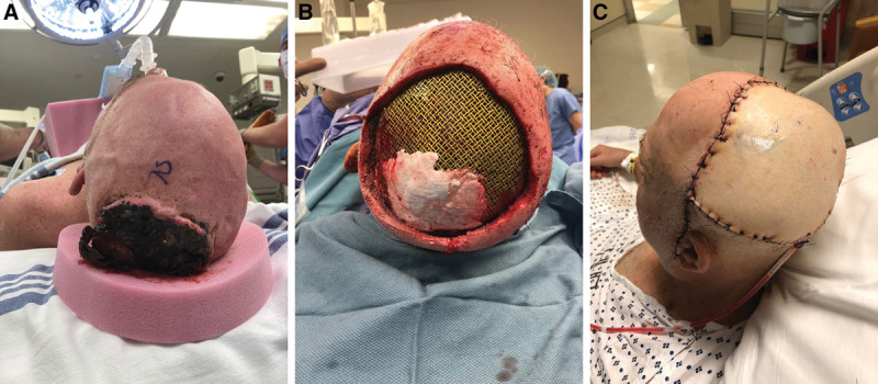
Fig. 1.

Initial patient presentation and surgery. A, Preoperative image demonstrating fungating scalp mass. B, Defect following excision of mass and titanium mesh cranioplasty. C, Postoperative image demonstrating ALT flap coverage of defect with a single drain in place.
