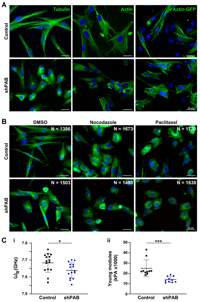
Figure 4.
Disrupted cytoskeletal spatial organization in shPAB human muscle cell cultures. A Representative images of control and shPAB human muscle cell cultures, stained with antibodies to tubulin and actin, and the actin filaments were visualized with actin-GFP. B Tubulin staining in control and shPAB myoblast cell cultures after DMSO, 100 nM nocodazole or 25 nM paclitaxel treatment for 2 h. Scale bar is 25 µm. C Measurements of cell-mechanics properties in control and shPAB cells using the Brillouin Light Scattering Microscopy (Ci) or the Atomic Force Microscopy (Cii). Measurements were carried out in myoblasts; every dot represents the median from 1000 measurements in a cell. Cell stiffness is measured by GHz, and the young modulus reports the cell surface tension. Averages and standard deviations are from N = 15 cells. Statistical significance was calculated with the student’s t-test.

