Figure 1.
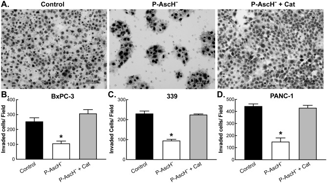
P-AscH− attenuates the invasive phenotype of PDAC in vitro. Cells were treated with P-AscH− or P-AscH− + catalase (200 U/mL) for 1 h then seeded at 1–3 × 10⁵ and incubated for 24 (PANC-1) or 48 h (BxPC-3 and 339). Data represent mean of invaded cells/field compared to control ± SE (n = 5, *p < 0.05; one-way ANOVA with Bonferroni’s multiple comparisons). (A) Representative invasion images from BxPC-3 PDAC cells in the presence of P-AscH− and/or catalase. (B) P-AscH− (2 mM for 1 h) decreased the percentage of invading BxPC-3 cells by 42% (255 +/− 23 cells vs 107 ± 15 cells). (C) P-AscH− (4 mM for 1 h) decreased the percentage of invading 339 patient derived PDAC cells by 41.5% (231 ± 11.5 cells vs 96 +/− 6 cells). (D) P-AscH− (4 mM for 1 h) decreased the percentage of invading Panc-1 PDAC cells by 34% (455 +/− 16.5 cells vs 151 +/− 30 cells).

