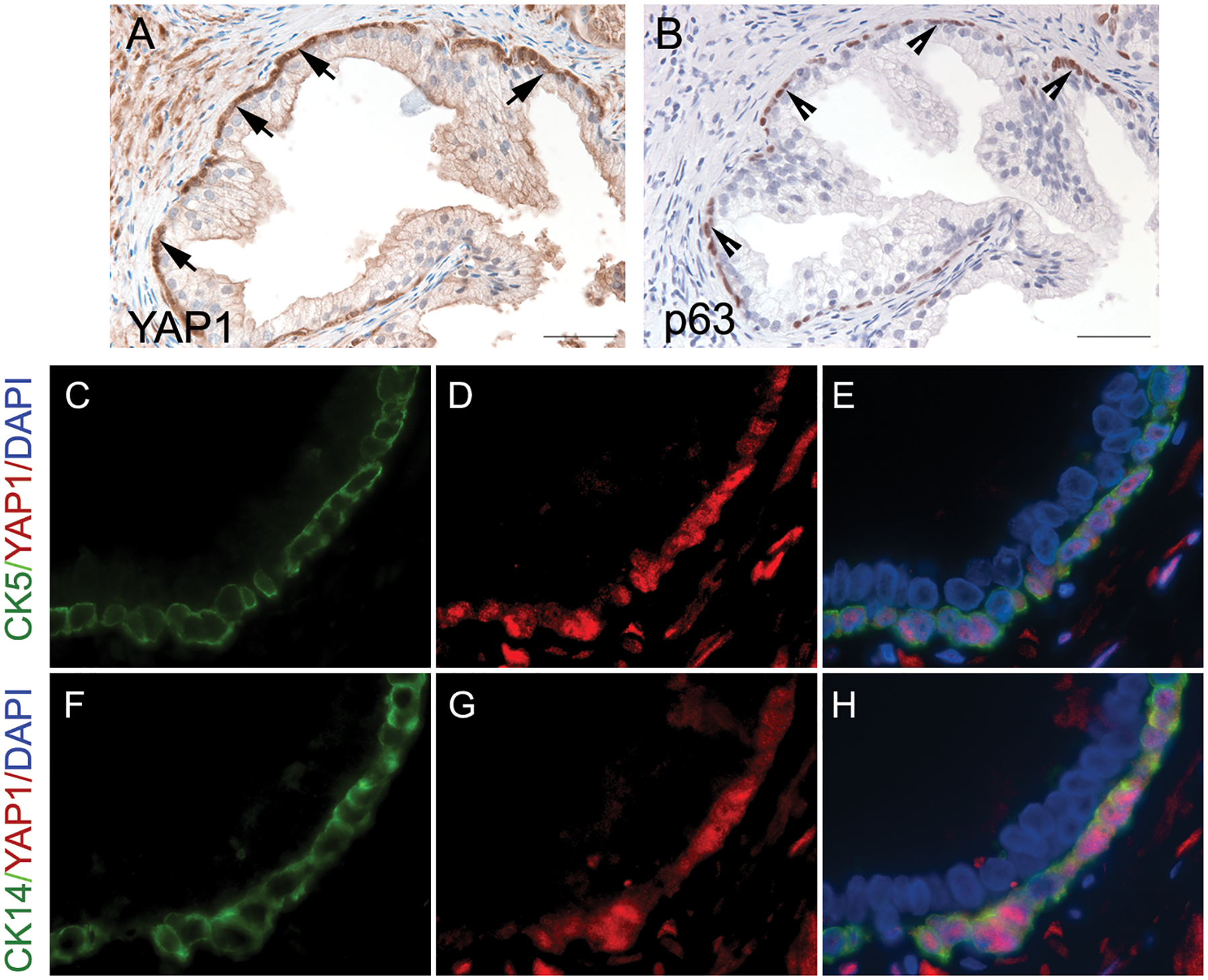
Fig. 1.

The expression of YAP1 in benign prostatic hyperplasia. (A) YAP1 displayed positive staining in basal cells (positive nuclear staining, indicated by the arrows) and negative staining in the prostatic luminal epithelial cells. (B) Immunohistochemical staining of p63 highlighted basal cells (indicated by the arrowheads) in the periphery of the prostatic gland. (C to H) Dual immunofluorescence staining of YAP1 and cytokeratin 5 (CK5) or cytokeratin 14 (CK14) confirmed the presence of YAP1 expression in prostatic basal epithelial cells.
