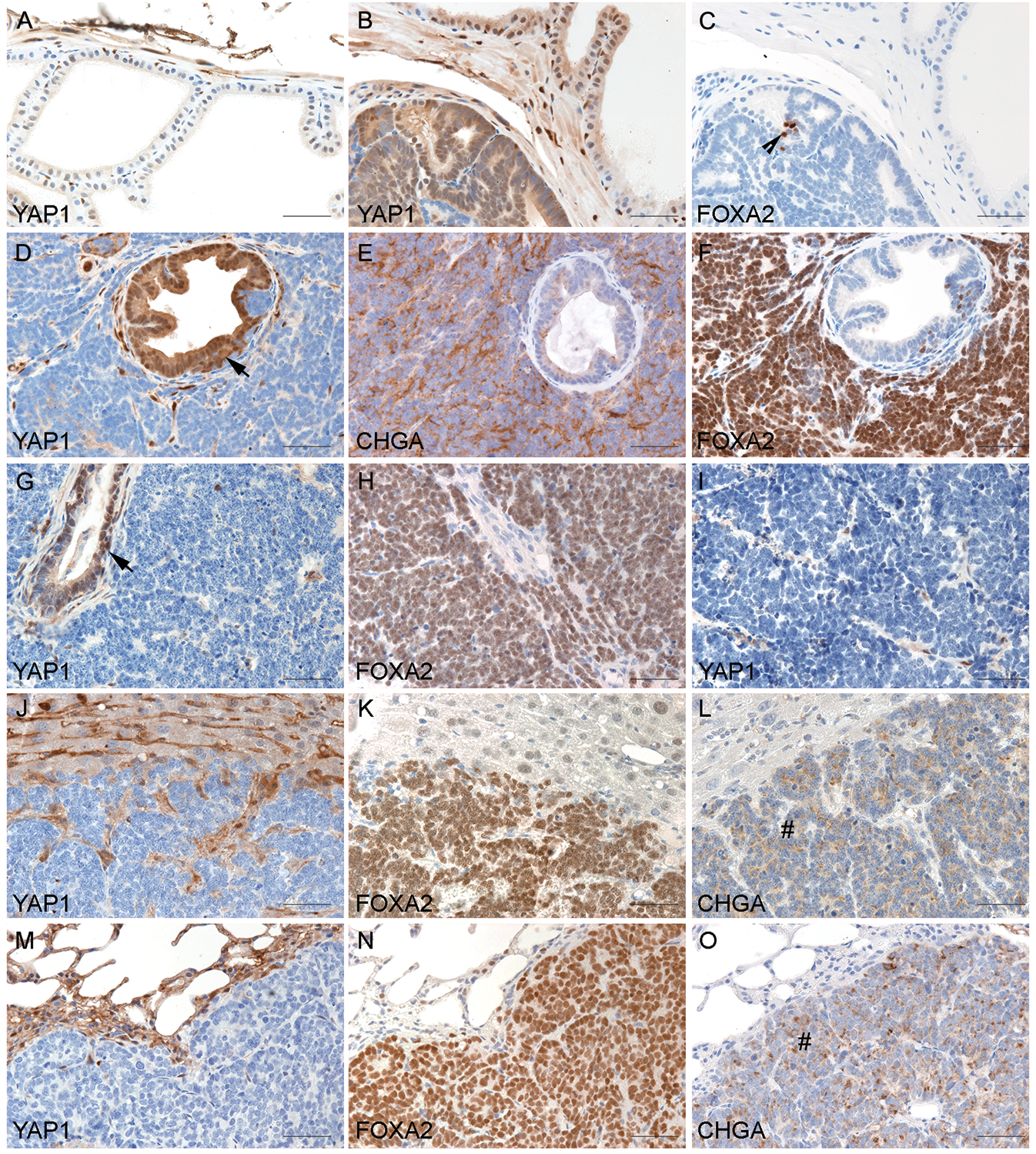
Fig. 2.

Microscopic examination of the prostate of TRAMP and LADY mice revealed that the expression of YAP1 increased in PIN but decreased in NEPC. (A) Wild-type prostate demonstrating positive staining of YAP1 in the occasional basal epithelia but negative staining in luminal epithelia. (B&C) Serial sections of a PIN tumor of TRAMP demonstrating diffuse positive staining of YAP1 and the rare FOXA2-positive cells in PIN (indicated by the arrowhead). (D to F) Serial sections of a NEPC tumor of TRAMP demonstrating diffuse positive staining of YAP1 in PIN components (indicated by the arrow) but negative in NEPC cells. NEPC markers chromogranin A (CHGA) and FOXA2 were stained positive in NEPC cells but negative in PIN. (G & H) Serial sections of a 12T-10 NEPC tumor demonstrating negativity of YAP1 in NEPC cells in contrast to the positive staining in focal PIN (indicated by the arrow). Positive staining of FOXA2 was seen in the NEPC cells. (I) NE10 tumor showed negative YAP1 staining. (J to O) The expression of YAP1 in NEPC metastases. Sections of the liver (J to L) and lung (M to O) with metastasis of NEPC demonstrated negative YAP1 staining in metastatic NEPC. In contrast, FOXA2 and CHGA were positive in NEPC metastases (indicated by the # signs).
