Fig. 3.

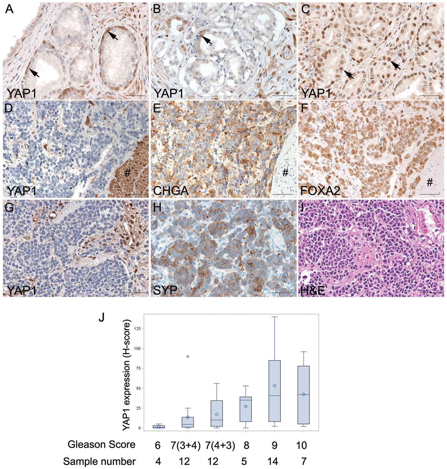
The expression of YAP1 in human prostatic tissues. (A) Benign prostatic tissue demonstrated positive YAP1 staining in the stroma cells and basal cells (indicated by the arrows). (B) Low-grade prostate adenocarcinoma. YAP1 expression was present in stromal cells and occasional basal cells (indicated by the arrow) but was absent in adenocarcinoma cells. (C) High-grade prostate adenocarcinoma. YAP1 was expressed in prostate adenocarcinomas (indicated by the arrows) with a Gleason score of 8 or higher. (D to F) Serial sections of a NEPC with focal adenocarcinoma. YAP1 protein was expressed in high-grade adenocarcinoma components (indicated by the # sign), but the expression was lost in NEPC cells. CHGA and FOXA2 were diffusely expressed in NEPC but not in adenocarcinoma cells. (G to I) Serial sections of a small cell carcinoma demonstrating negative YAP1 and positive synaptophysin (SYP) staining in NEPC cells. (J) Distribution of YAP1 expression (H-score) among PCa with various Gleason grade. The expression of YAP1 was associated with Gleason score (Spearman correlation test, r=0.4962, p<0.0001).
