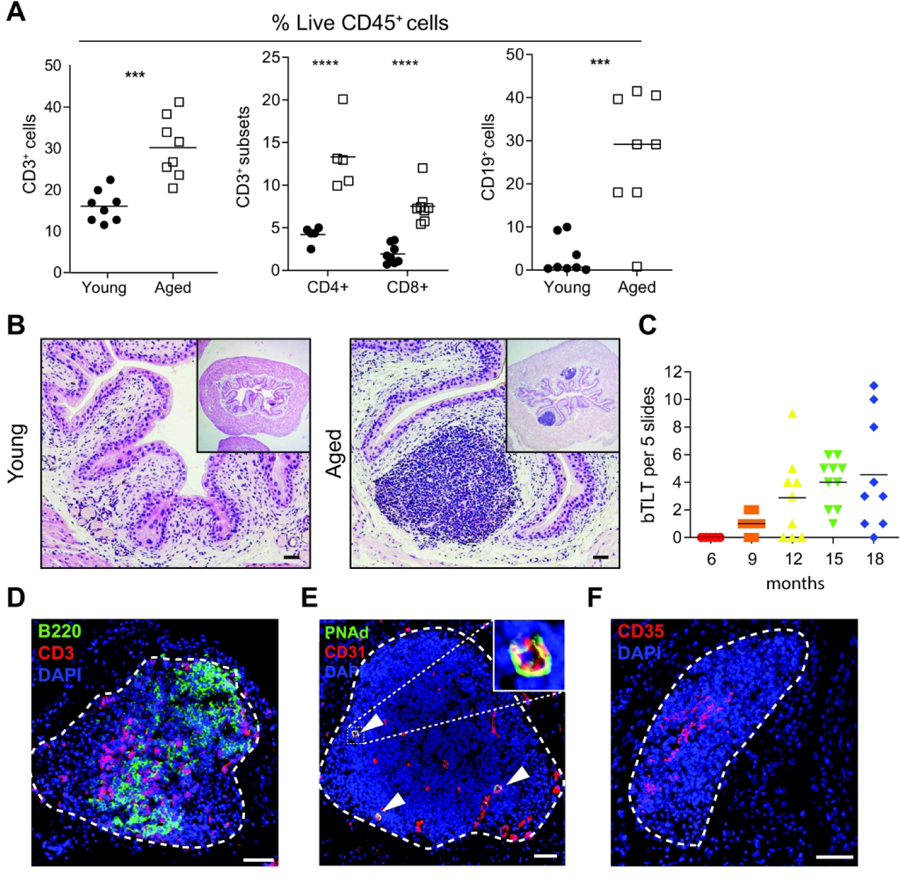
Figure 3. Lymphoid infiltrates form bladder tertiary lymphoid tissues (bTLT) during aging.

(A) Frequency of B cells, T cells, and T cell subsets among live CD45+ cells in young and aged bladders by flow cytometry. n=5–8 per group. (B) Representative H&E images of young and aged bladders. (C) Number of bTLT over the life course of mice n=5–10/group. (C) Representative image of B cells (B220+, green) and T cells (CD3+, red) in segregated zones within bTLT in aged mice. (D) Representative image of CD31+(red) PNAd+(green) high endothelial venules (white arrowheads) within bTLT in aged mice. (E) Representative image of CD35hi follicular dendritic cell network within bTLT in aged mice. All nuclei stained with DAPI (blue). All scale bars, 50 μm.
