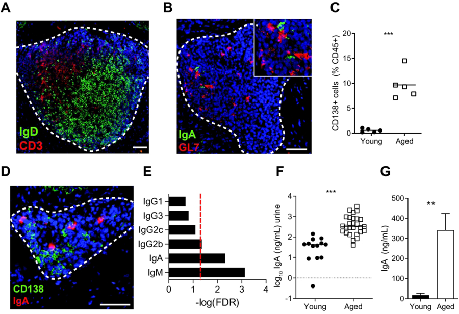
Figure 4. bTLT are centers for B cell recruitment, activation, germinal center reactions, and plasma cell differentiation.

(A) Representative image of naive B cells (IgD+, green) and T cells (CD3+, red) within bTLT of aged bladders. (B) Representative image of IgA+ cells within a GL7+ (green) germinal center of a bTLT. (C) Frequency of live CD45+CD138+ plasma cells in young and aged bladders by flow cytometry. n=5/group. (D) Representative image of IgA+CD138+ plasma cells within bTLT of aged bladders. (E) FDR -adjusted P values of IgM and class-switched isotypes from tissue RNA-seq of young and aged bladders. Red line, p=0.05. (F) Concentration of IgA in urine of young (n=13) and aged (n=27) mice. (G) Concentration of IgA in supernatants of young and bladders cultured ex vivo for 24 hours. n=5/group. All scale bars, 50 μm. **p<0.01, ***p<0.001. Mann-Whitney U test.
