
Fig. 2. Dara induces direct CD38+ NK cell activation.
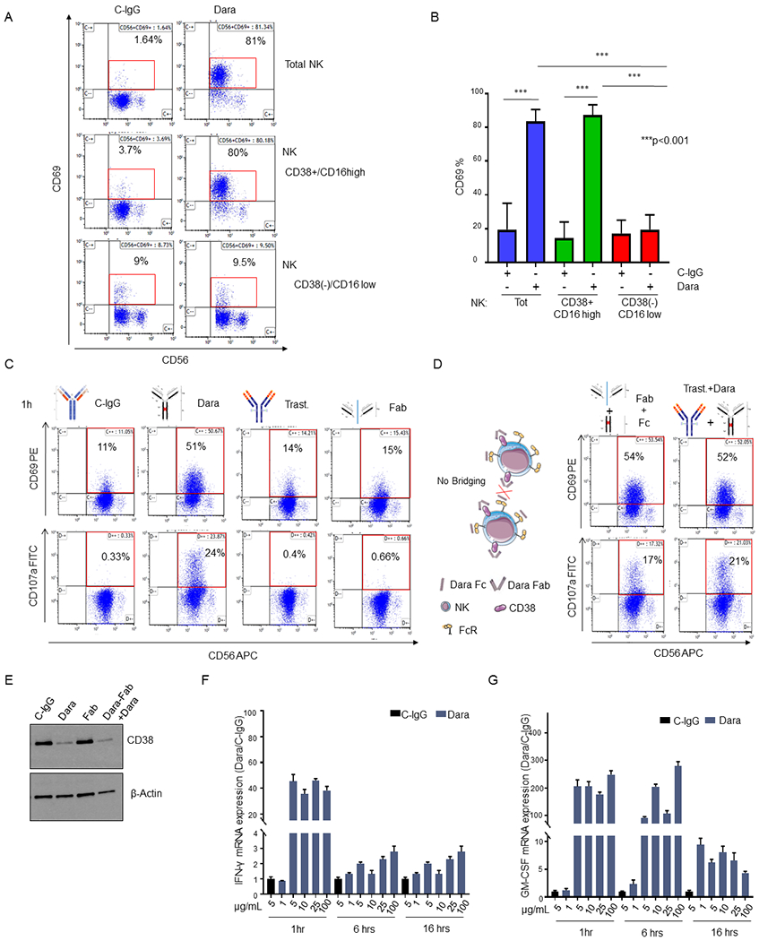
A) Representative flow analysis of CD69 on total NK, CD38+NK and CD38(−) NK cell fractions isolated from a healthy donor upon overnight Dara treatment; B) Bar graph representing the median ±SD of CD69 expression in total, CD38(−) and CD38+ NK cells treated with Dara or C-IgG (p<0.001). The experiment was repeated using three healthy donors in triplicate; C,D) Flow cytometry analysis of CD69 (top panels) and CD107a (bottom panels) in total NK cells treated for 1hr with C-IgG (C), Dara (C), Trast (C), Dara Fab (Fab) (C), Dara Fab + Dara Fc (Fab+Fc) (D), or Trast+Dara (D) at 10 μg/mL for 1 hr using the same HD (n=1) for each treatment and internal controls; D) Graphical Illustration (left panel) showing the fully saturated CD38 on NK cells after the addition of the purified Dara Fc fragment for 30 min, which followed the addition of Dara Fab, excluding a possible bridging between NK cells; E) Western Blot analysis showing CD38 protein levels in NK cells treated with either C-IgG (10 μg/mL), Dara (10 μg/mL), Dara-Fab or Dara-Fab+Dara for 48 hrs. The experiment was run in duplicate; F, G) IFN-γ and GM-CSF mRNA expression levels in NK cells under Dara treatment at different concentrations (from 1 μg/ml to 100 μg/ml) and different time points (1-16 hrs) compared to C-IgG; GADPH mRNA was used for normalization.