
Fig. 5. Dara-resistant patients retain surface CD38 on MM cells.
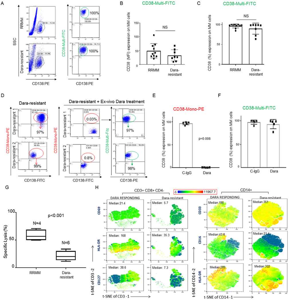
A) Representative flow analysis of CD38 expression showing no significant modulation of CD38 expression (%) on CD138+ MM-cells obtained from Dara-resistant (n=8) compared to RRMM patients (n=10); B,C) Bar graphs showing CD38 surface expression by MFI (B) and % (C) among Dara-resistant and RRMM patients. The Mann-Whitney-Wilcoxon test was performed; D) Representative flow analysis of CD38 on CD138+ MM cells of two Dara-resistant patients where BMCs were treated ex vivo with Dara for 1 hr, showing lack of CD38 in MM cells treated with Dara and stained with CD38-Mono PE, and presence of CD38 when the same cells were stained with CD38-Multi FITC; E,F) Bar graphs showing CD38 surface levels in CD138+ MM cells of 5 Dara-resistant patients after ex-vivo Dara incubation and stained with CD38-Mono PE (E) and CD38-Multi FITC antibodies (F); G) Bar graph showing killing induction (%) assessed by flow based assay in a set of RRMM patients (n=4) and Dara-resistant patients (n=6) treated with Dara ex vivo. Percentage of dead cells was calculated by gating in 7-AAD positive cells among target cells (MM1.S GFP+ cells); H) tSNE heatmap statistic plot obtained by CyTOF analysis showing CD69, HLA-DR and CD127 expression (Median) in the CD8+ cell subpopulation (gated in total CD3+ cells) of PBMCs isolated from Dara-resistant and Dara-responding patients and tSNE heatmap statistic plot obtained by CyTOF analysis showing CD33 and CD16 expression (Median) of PBMCs isolated from a representative Dara-resistant and Dara-responding patients.