
Figure 3.
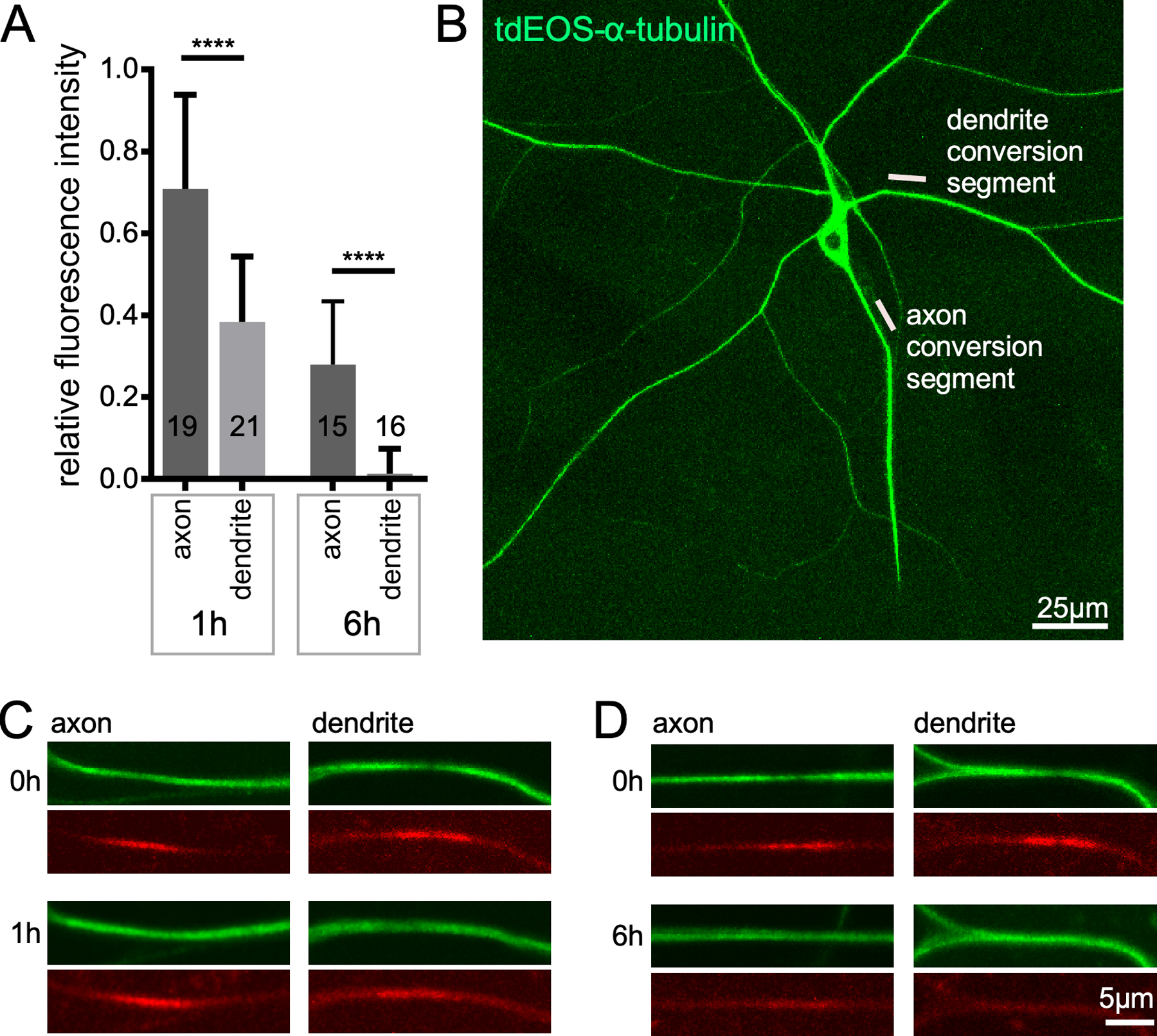
Microtubules turn over faster in dendrites than axons. Photoconvertible tdEOS-α-tubulin (111) was expressed in Drosophila class IV dendritic arborization neurons with 477-Gal4. Whole larvae were mounted for live imaging; an example image of the ddaC neurons used is shown in B. Ten micron regions in either the dendrite or axon were converted from green to red using UV light. Example images of converted segments can be seen in red channel of the 0h images in C and D. The green unconverted signal is also reduced in the conversion region. 1h after conversion the converted region can still be seen in axons and dendrites (C). However, by 6h only the axon still has a visible conversion region. The conversion signal was set at 1 in the 0h images, and the relative amount remaining at 1h and 6h is shown in A. The error bars show standard deviation and the number on the bars are the numbers of animals analyzed. Dendrite data is a re-analysis of data from (52). The statistical test used was a t-test, ****p<0.0001.