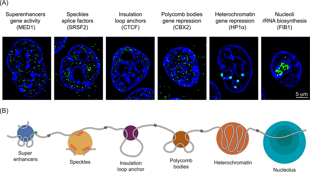
Figure 1. Biomolecular Condensates in the Nucleus.
(A) Structured illumination microscopy images of immunofluorescence for the protein indicated in parentheses in murine embryonic stem cells. Immunofluorescence for indicated protein is colored green, and signal from Hoechst, a DNA stain, is colored dark blue (unpublished results AD and RAY). Condensates are denoted by their name (e.g., superenhancers), their function (e.g., gene activity), and the protein that provides the immunofluorescent signal (e.g., MED1). (B) Cartoon depiction of how various nuclear condensates organize and are organized by different chromatin substrates. The grey line represents the chromatin fiber, green arrow designates active transcription start site, and red squiggled lines represent RNA. For a more complete list of nuclear condensates see Table 1.
Abbreviations: CBX2, chromobox protein homolog 2; CTCF, CCCTC-binding factor; HP1α, heterochromatin protein 1α.

