
Figure 4. Microglia depletion increases neuronal injury in response to KA treatment.
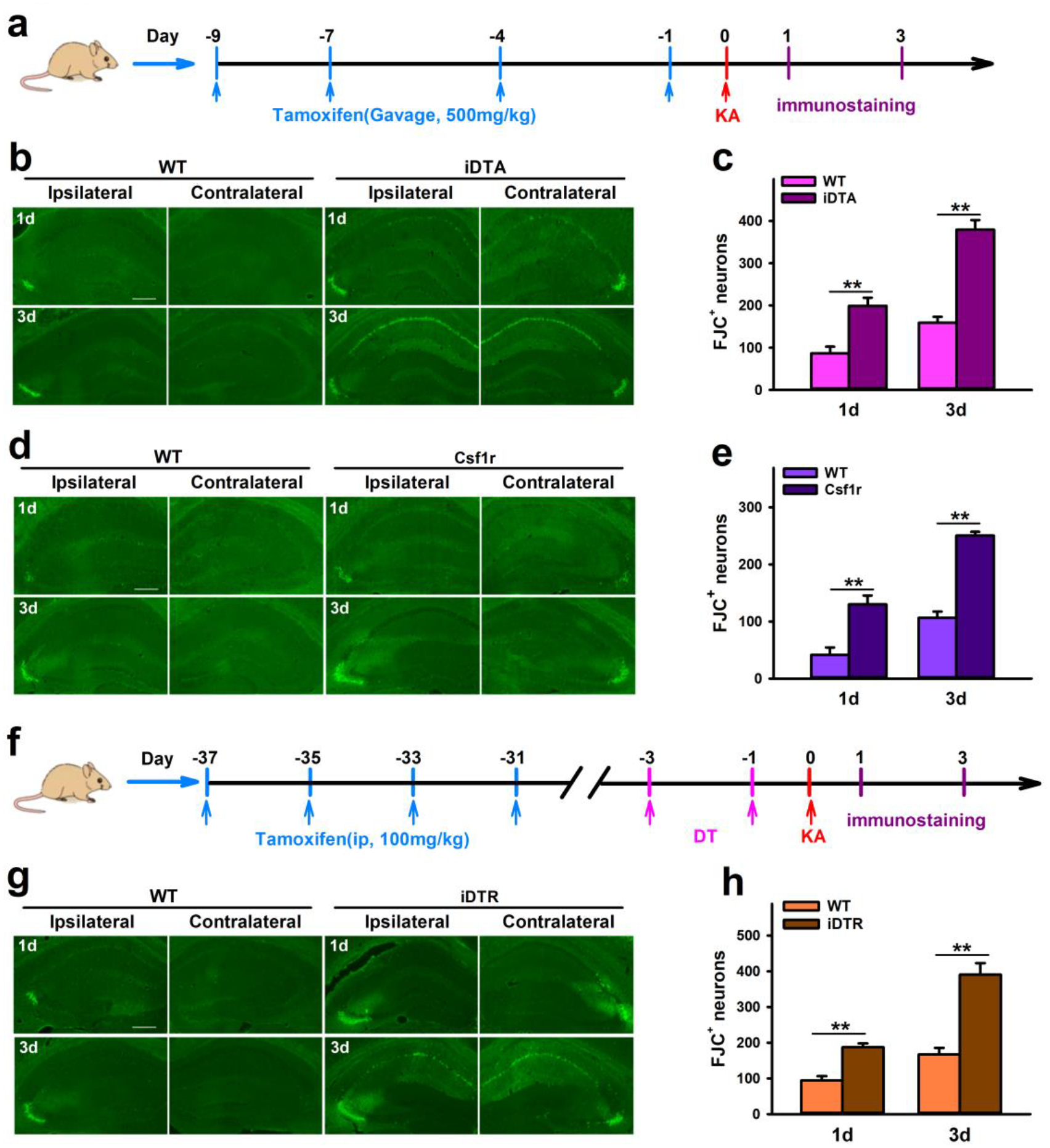
a, Drug treatment and immunostaining scheme in iDTA and Csf1r mice. b–e, Representative images and quantification of FJC+ neurons show that CX3CR1+ cell depletion worsens neuronal damage in the hippocampus of iDTA mice (b, c) and Csf1r mice (d, e) 1d and 3d after ICV KA treatment. f, Drug treatment and immunostaining scheme in iDTR mice. g–h, Representative images and quantification of FJC+ neurons show that specific microglia depletion worsens neuronal damage in the hippocampus of iDTR mice 1d and 3d after KA treatment. Scale bar=100 μm. Data presented as means ± SEM, n= 3, **P<0.01 vs control group.