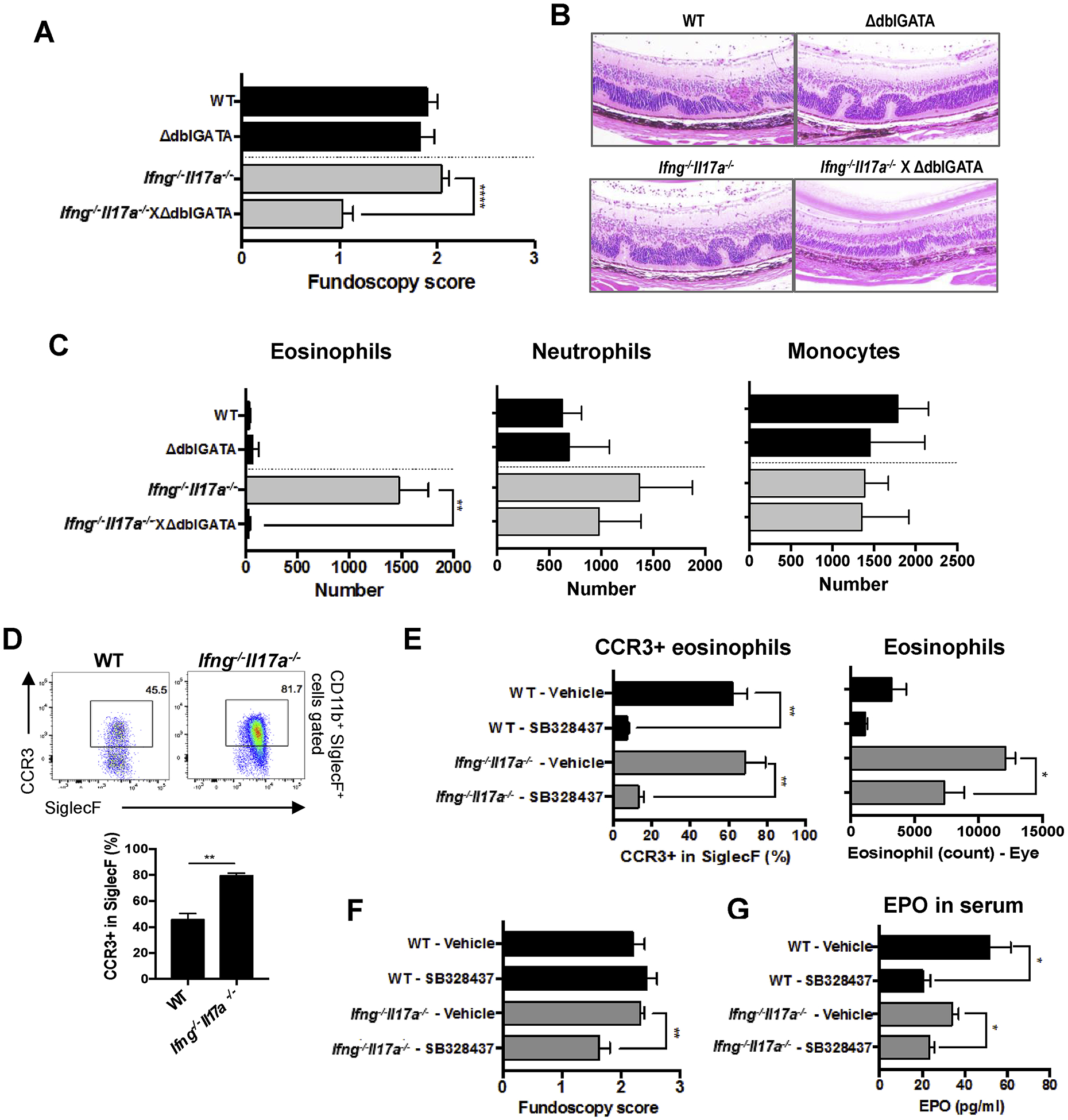
Fig. 2.

Eosinophils contribute to the pathogenesis of EAU in the absence of IFN-γ and IL-17A. (A–C) WT, ΔdblGATA, Ifng−/−Il17a−/−, Ifng−/−Il17a−/−ΔdblGATA mice on C57BL/6 background were immunized with 100 μg of p651–670. (A, B) EAU scores were evaluated by (A) fundoscopy, and (B) histology with H&E stain. Original mag, 20x. (C) The number of SiglecF+, Ly6C−Ly6G+, or Ly6C+Ly6G− cells in the CD45+CD11b+CD11c− gates from eyes on day 16. (D) The frequency of CCR3+ cells in eye-infiltrating eosinophils was determined by FACS. (E, F, G) WT and Ifng−/−Il17a−/− mice were s.c. treated with 200 μg of a CCR3 antagonist (SB328437) every day from one day before immunization. (E) The frequency of CCR3+ eosinophils and total eosinophils in eye-infiltrating cells were determined by FACS. (F) Fundoscopy score on day 15 days after immunization. (G) Levels of Eosinophil Peroxidase (EPO) in the serum were measured by LEGENDplex on day 16. (A) Data combined from 3 experiments (WT; n = 14, ΔdblGATA; n = 10, Ifng−/−Il17a−/−; n = 14, Ifng−/−Il17a−/− × ΔdblGATA; n = 5). (B–G) Representative data from 2 independent experiments, and each group contains at least 3 mice. Data shown as mean ± SEM. Significance was determined using Mann-Whitney U test (A, F), or unpaired t-test (C, D, E, G). *p < 0.05, **p < 0.01.
