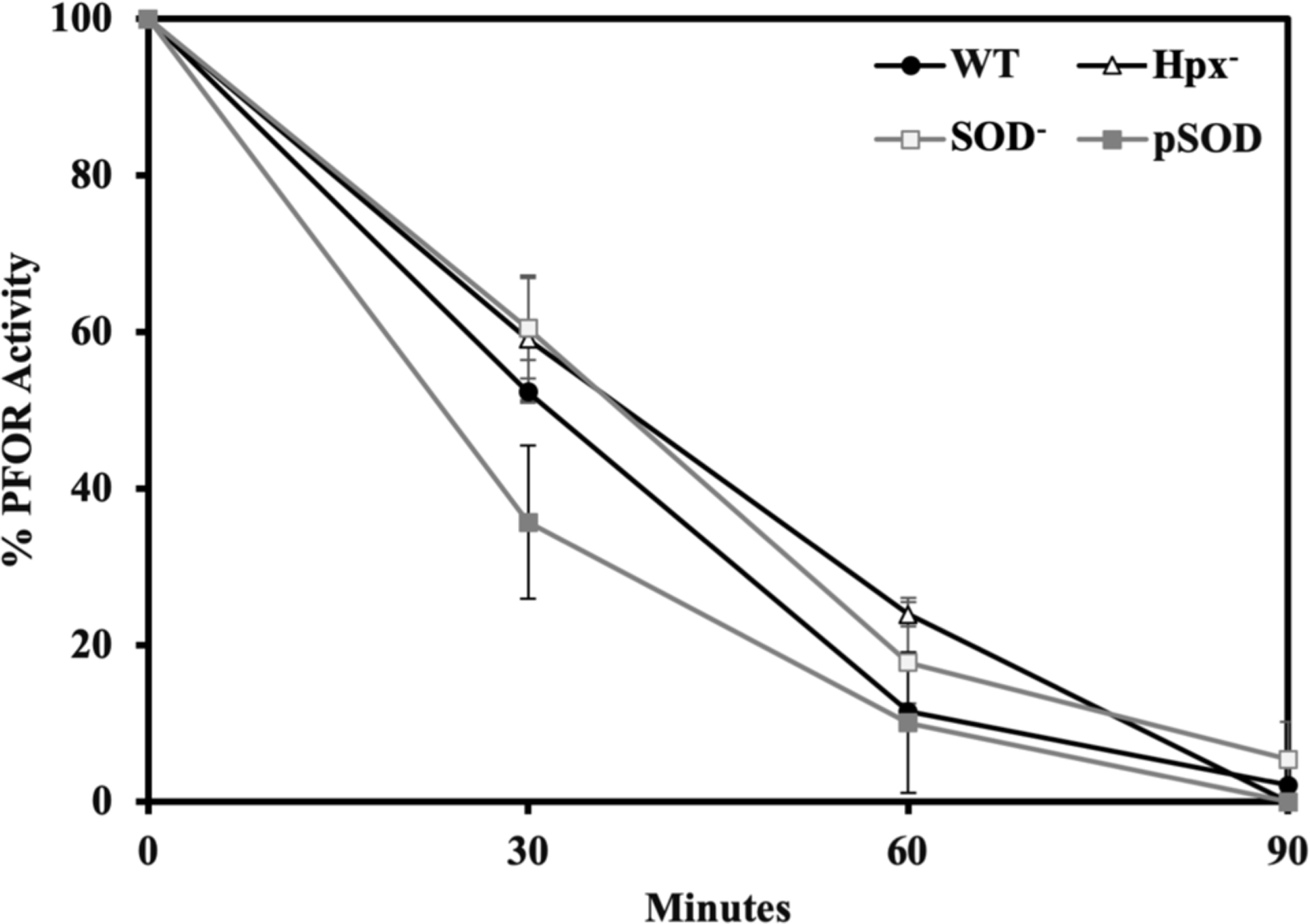
Figure 5. The rate of PFOR inactivation in aerated cells is independent of the intracellular concentrations of H2O2 and superoxide.

Cells growing exponentially in anoxic BHIS media were washed, resuspended in glucose buffer containing chloramphenicol, and aerated. At intervals aliquots were returned to the anaerobic chamber and PFOR activity was assayed. Strains: WT (BT5482), Hpx− (SM135), SOD− (LZ01), and pSOD (LZ200). Error bars and values after ± represent the standard error of the mean of three biological replicates.
