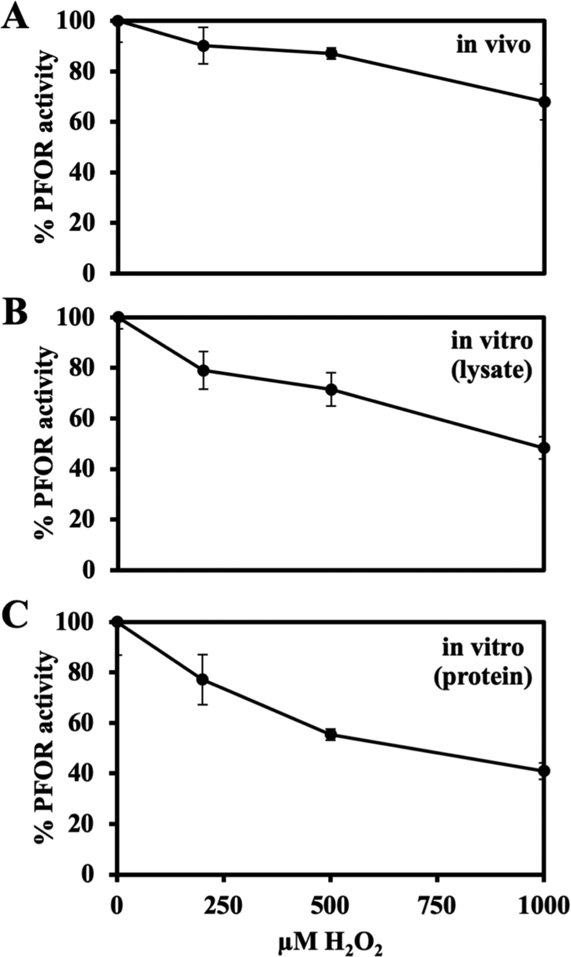
Figure 6. Physiological (<10 μM) concentrations of H2O2 cannot inactivate purified PFOR.

All cell growth, protein purification and enzyme assays were performed in the anaerobic chamber; see Materials & Methods for details. (A) Exponentially growing Hpx− (SM135) cells were washed with and resuspended in Tris buffer and incubated with H2O2 for 15 minutes at 4 oC.. PFOR activity was then assayed. (B) In vitro: Cell lysates were prepared from Hpx− cells. Lysates were incubated with H2O2 for 15 minutes at 4 °C, and PFOR activity was assayed. (C) The purified B. thetaiotaomicron PFOR was incubated with H2O2 for 15 minutes at 4 °C and then assayed. Error bars represent the standard error of the mean of three biological replicates.
