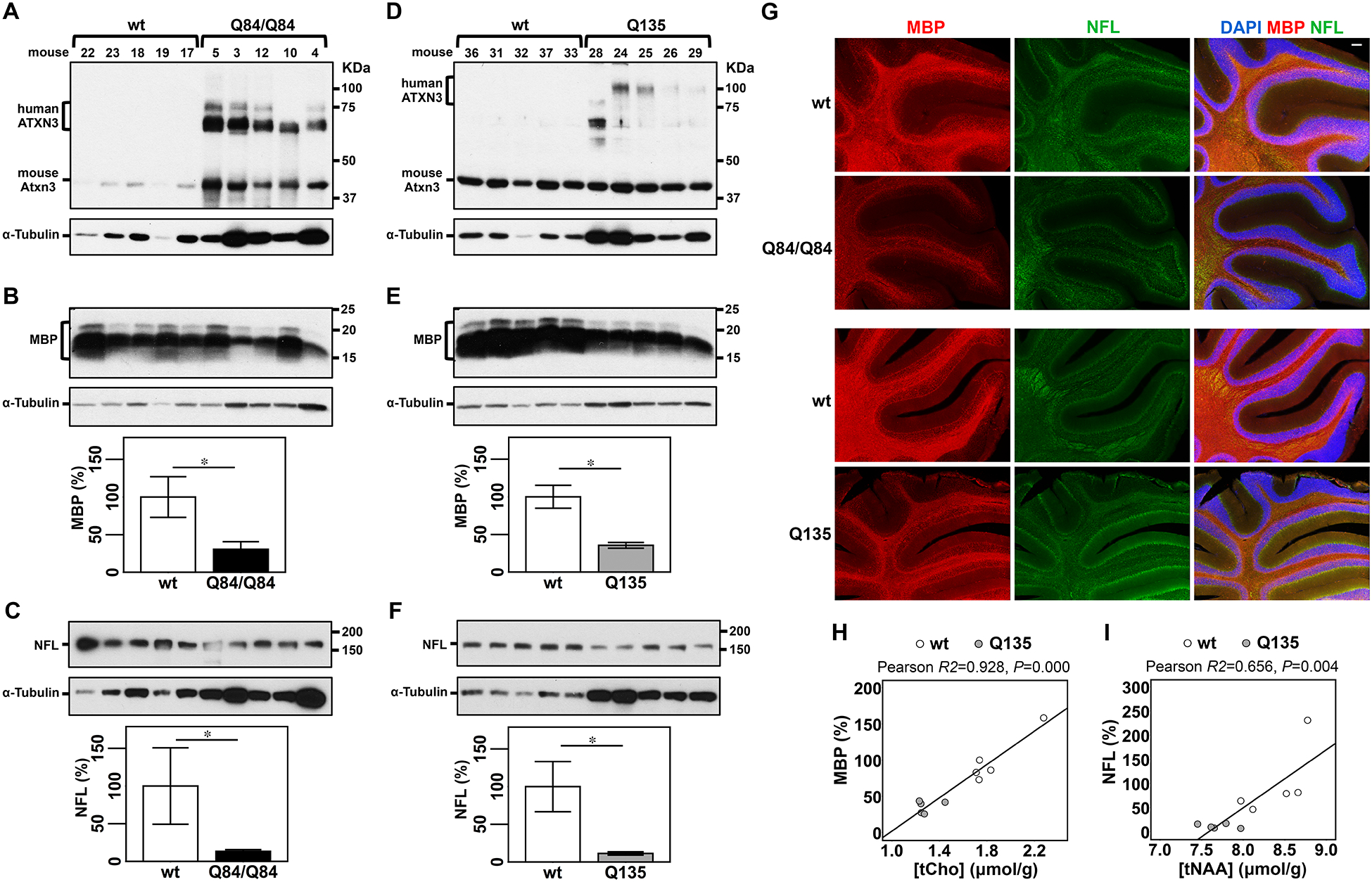
Figure 3: Cerebella of SCA3 transgenic mice show decreased levels of myelin basic protein (MBP) and neurofilament medium (NFL) that correlate with concentrations of tCho and tNAA, respectively, in Q135 mice.

A,D) Anti-ATXN3 immunoblot (anti-MJD antibody) detecting mutant human ATXN3 and endogenous mouse Atxn3 in cerebellar soluble protein extracts of the subset of Q84/Q84 (A), Q135 (D) and corresponding littermate wild type mice used to assess levels of MBP (B,E) and NFL medium (NFL) (C,F). Western blots show decreased levels of MBP and NFL in cerebella of Q84/Q84 (B,C) and Q135 (E,F) compared with controls. Graphs show quantification of protein bands by densitometry. Bars represent the average percentage of protein relative to respective wild type controls, normalized for a-Tubulin (± SEM). Statistical significance determined by Student’s t-test is indicated as *P<0.05. G) Representative images of immunofluorescent labelling of MBP (red) and NFL (green) show decreased signal of these two proteins in cerebella of Q84/Q84 and Q135 mice compared with corresponding litter mates (N=4 mice per group). Nuclei were labeled with DAPI (blue). Scale bar, 100 μm. H, I) Plots showing Pearson correlations of levels of MBP with tCho (H), and NFL with tNAA (I) in Q135 (grey circles), and their respective wt littermate mice (white circles).
