
Figure 4: Cerebellar lobules of patients with SCA3 show decreased levels of MBP and NFL medium and demyelination.
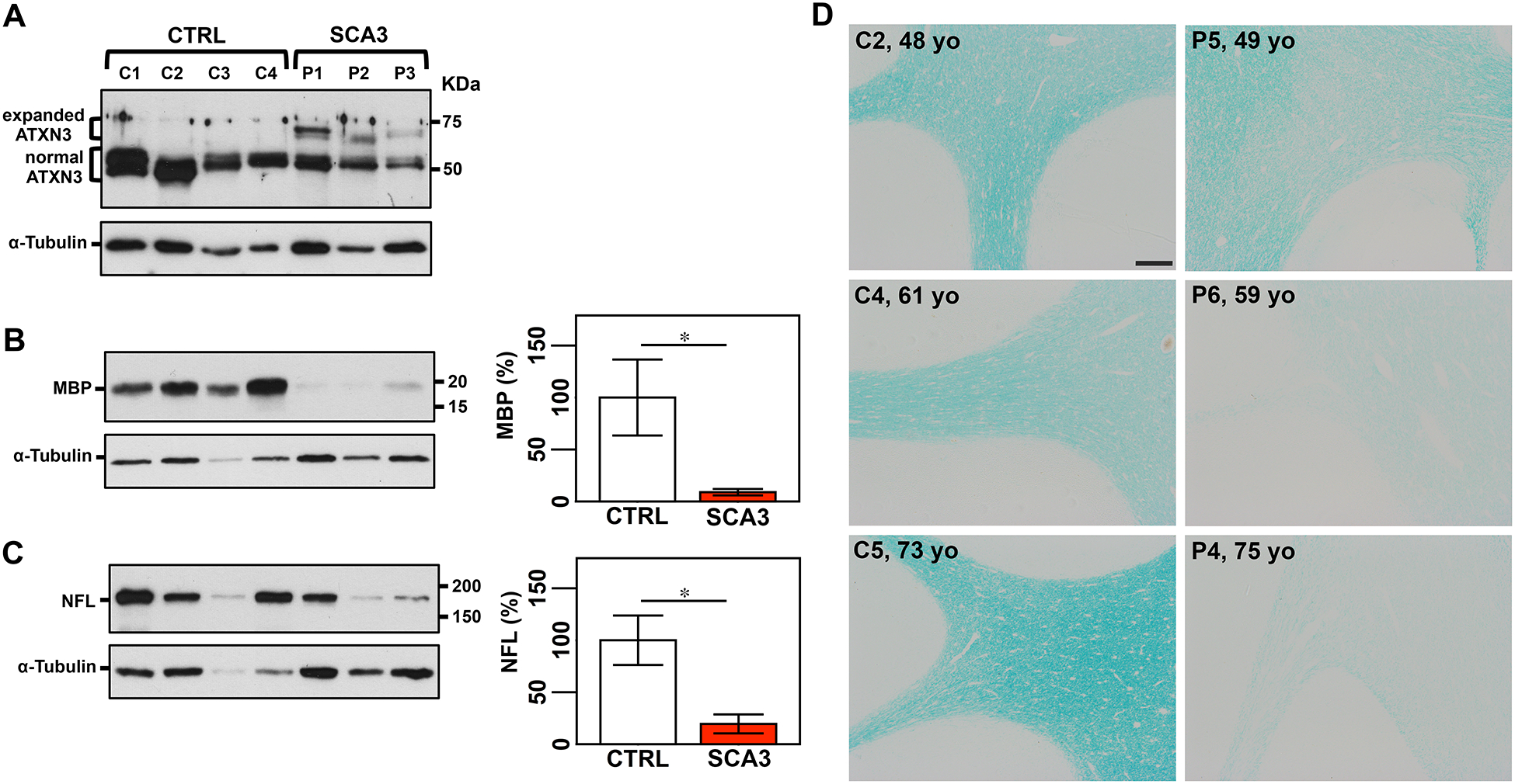
A) Anti-ATXN3 immunoblot (anti-MJD antibody) detecting expanded and normal ATXN3 in soluble protein extracts from cerebellar cortex of patients with SCA3 and control individuals. Detection of MBP (B) and NFL medium (C) by Western blotting shows lower levels of both proteins in patients compared with controls. Graphs show quantification of protein bands by densitometry. Bars represent the average percentage of protein relative to respective wild type controls, normalized for a-Tubulin (± SEM). Statistical significance determined by Student’s t-test is indicated as *P<0.05. D) Luxol fast blue staining of cerebellar sections shows signs of demyelination in patients with SCA3 compared with aged-matched unaffected individuals, which is more evident in older patients. Scale bar, 200 μm.