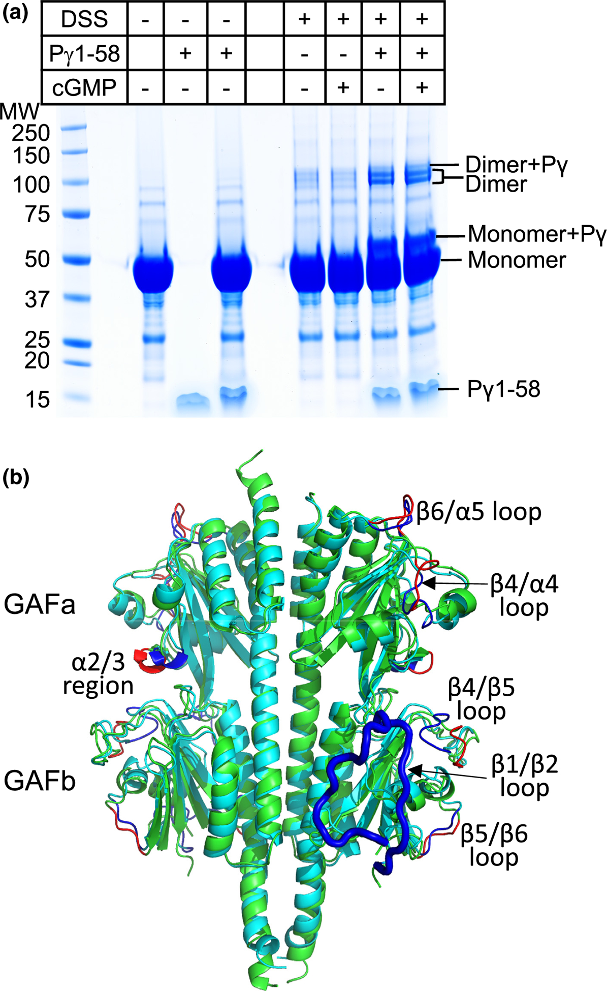
Figure 3.

Structural model of the apo state of cone PDE6 GAFab determined by chemical cross-linking and mass spectrometry. (a) SDS-PAGE of a cross-linking experiment in which 12 μM GAFab was incubated in the presence or absence of cGMP (10-fold molar excess) and/or 120 μM Pγ1–58 prior to addition of a 50-fold molar excess of the chemical cross-linker BS3. (b) Structural alignment of cross-linked refined apo-GAFab (cyan) with the x-ray structure (green), with major differences indicated for the x-ray (red) and apo (dark blue) structures; the GAFb β1/β2 loop (residues 286 to 310) that is missing in the x-ray structure is highlighted as a thick blue loop in the apo structure.
