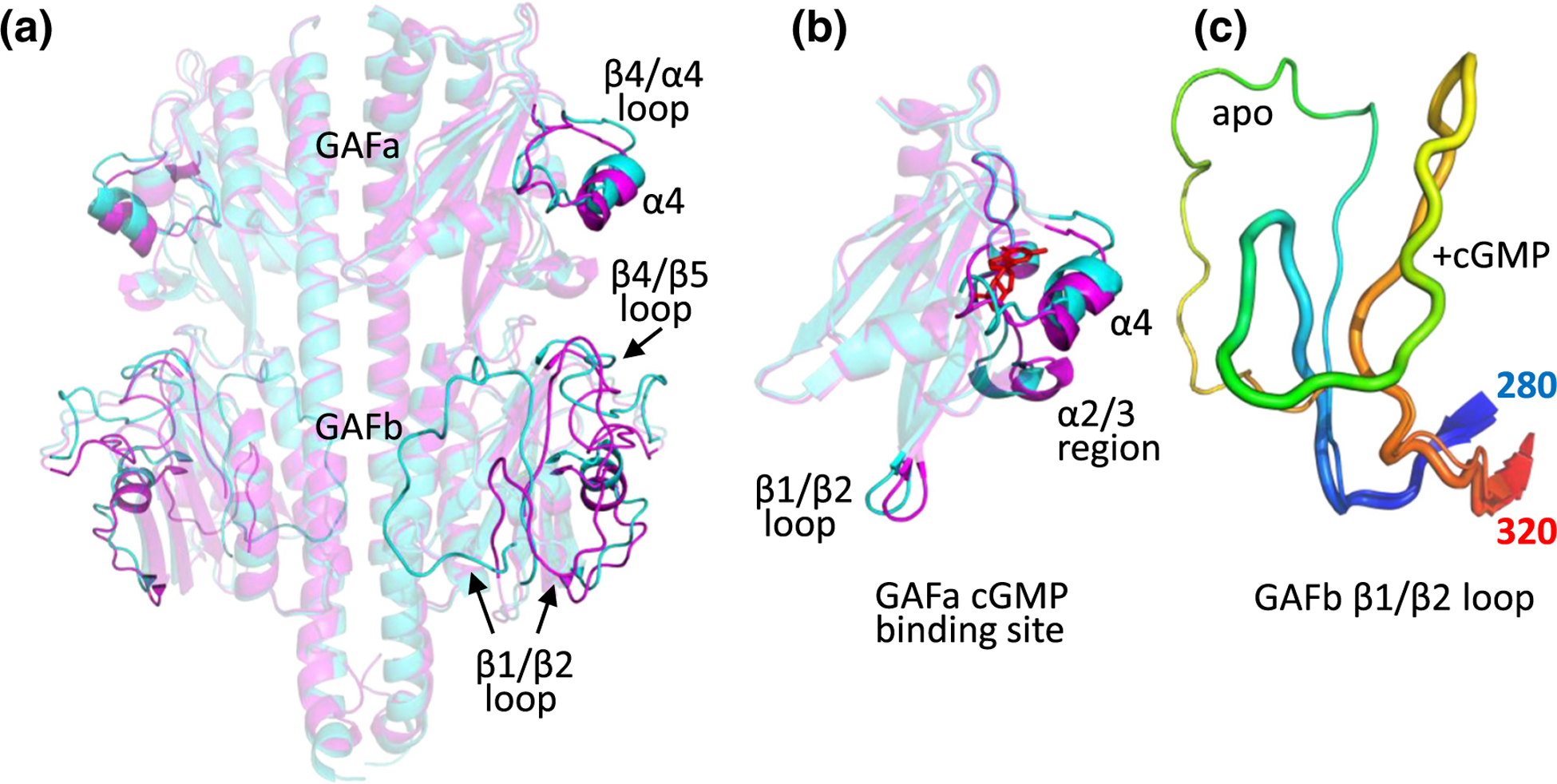
Figure 4.

Structural changes in PDE6 GAFab upon binding of cGMP. (a) Comparison of apo GAFab (cyan) with GAFab·cGMP (magenta) with significant conformational differences highlighted. (b) Comparison of the conformational states of the GAFa domain in its apo (cyan) and cGMP-bound state (magenta, with cGMP (red) docked). (c) Comparison of the conformation of the GAFb β1/β2 loop (residues 280 (blue) to 320 (red)) in the apo (thin loop) and GAFab·cGMP (thick loop) states.
