
Figure 1. Diabetic ZDF rats have reduced PNN structures in the arcuate nucleus.
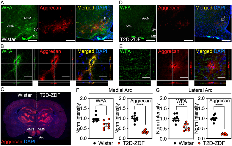
Immunofluorescent detection of WFA, a lectin that binds to the CS-GAG glycan component of PNNs1, and aggrecan, a PNN-associated CSPG core protein8, in coronal sections of rat hypothalamus (30 μm) from (A-C) normoglycemic Wistar controls and (C-E) age-matched, hyperglycemic ZDF rats. (A, D) Low-magnification view of WFA+ / aggrecan+ PNNs throughout the ArcM and ArcL areas of the mediobasal hypothalamus. Scale bar: 200 μm. (B, E) Higher-magnification orthogonal views of WFA+ / aggrecan+ PNN enmeshments. Scale bar: 25 μm. (C) Tiled panoramic image of aggrecan from a coronal section from a (left) normoglycemic Wistar control and a (right) age-matched diabetic ZDF rat shows selective loss of PNN labeling in the Arc of ZDF rats. Scale bar: 1 mm. Quantification of the mean fluorescence intensity of PNNs averaged from medial hypothalamic sections (−2.2 to −2.8 mm posterior from bregma) for (F) medial Arc and (G) lateral Arc areas from normoglycemic Wistar and diabetic ZDF rats and normalized to the Wistar mean fluorescence intensity averages (n=8 rats/group; mean ± SEM). **P < 0.01, ***P < 0.001, ****P < 0.0001 versus Wistar controls; Student’s t-test (unpaired, two-sided); (F) WFA-ArcM p=0.0.0036, aggrecan-ArcM p=<0.0001, WFA-ArcL p=0.0004, aggrecan-ArcL p=<0.0001. Two separate cohorts of diabetic ZDF and normoglycemic Wistar rats were analyzed, and the results were reproducible between studies. Arc, arcuate nucleus; ArcL, lateral Arc; ArcM, medial Arc; ME, median eminence; VMN, ventromedial nucleus; 3V, 3rd ventricle.