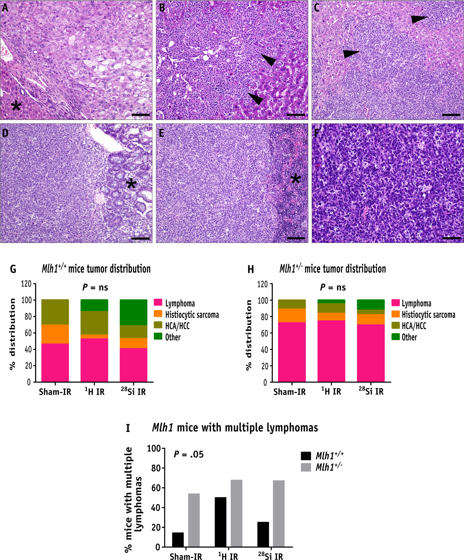
Fig. 2.

Hematoxylin and eosin analysis of tumors obtained from Mlh1+/+ and Mlh1+/− mice. (A) Primary hepatocellular carcinoma composed of solid plates and lobules of poorly differentiated hepatocytes (asterisk denotes normal liver, bar = 50um). (B) Histiocytic sarcoma in the liver, composed of infiltrative sheets of pleomorphic round cells effacing normal hepatic parenchyma (arrowheads, bar = 50um). (C) Lymphoma in the liver, composed of sheets of neoplastic lymphocytes (arrowheads) infiltrating and effacing normal hepatic parenchyma (arrowheads, bar = 50um). (D) GALT lymphoma is expanding the submucosa of the small intestine and infiltrating the overlying intestinal mucosa (asterisk denotes normal mucosa, bar = 50um). (E) Lymphoma effacing the spleen (asterisk denotes normal spleen, bar = 50um) and (F) mesenteric lymph node (bar = 20um), composed of sheets of atypical lymphocytes. (G) Percent distribution of types of tumors arising in Mlh1+/+ and (H) Mlh1+/− mice post sham, 1H ion, or 28Si ion irradiation. (I) The bar graph represents the percentage of mice with multiple lymphomas in Mlh1+/+ and Mlh1+/− cohorts post radiation exposure. Histopathology was performed on 13 to 32 tumors of Mlh1+/+ origin and 18 to 56 tumors of Mlh1+/− origin. Tumor distribution was analyzed by the χ2 test, and multiple lymphoma incidence was analyzed by 2-way analysis of variance. Abbreviation: GALT = gut associated lymphoid tissue; HCA = hepatocellular adenoma; HCC = hepatocellular carcinoma; ns = nonsignificant.
