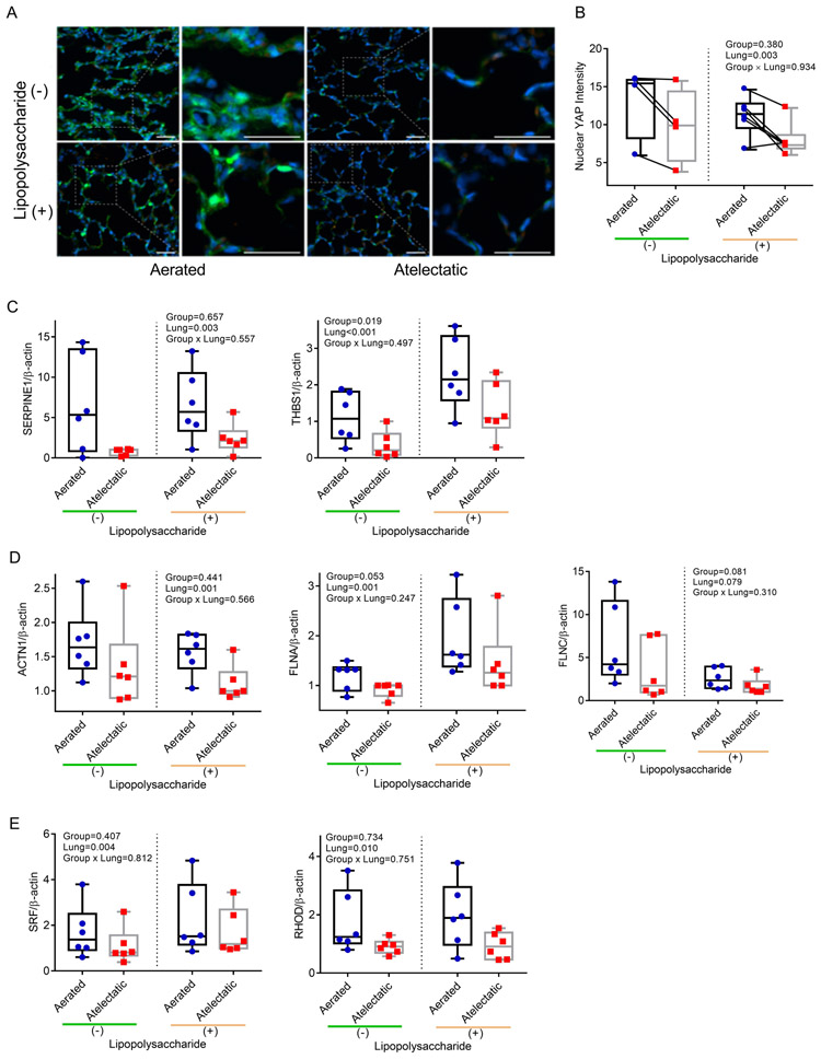
Figure 9. YAP is involved in atelectasis-associated barrier dysfunction.
(A) Representative images for YAP (green) from aerated and atelectatic regions in LPS(−) (n=6 animals) or LPS(+) (n=4 animals). Nuclei were stained with Hoechst in blue and autofluorescent blood cells appear in red; scale bars represent 20 μm. (B) Nuclear YAP intensity was markedly reduced in atelectatic vs aerated regions. Each connected dot pair represents atelectatic and aerated regions from a single animal. (C-E) Real-time polymerase chain reaction validation documenting lower mRNA expression for YAP-responsive genes SERPINE1 and THBS1 (C), cytoskeleton-associated proteins ACTN1, FLNA and FLNC (D) and actin dynamics associated factors SRF and RHOD (E) in atelectatic than in aerated regions (n=6 animals). P-values corresponding to analyzed effects (LPS exposure (Group), lung region (Lung), and their interaction (Group × Lung)) are indicated.

