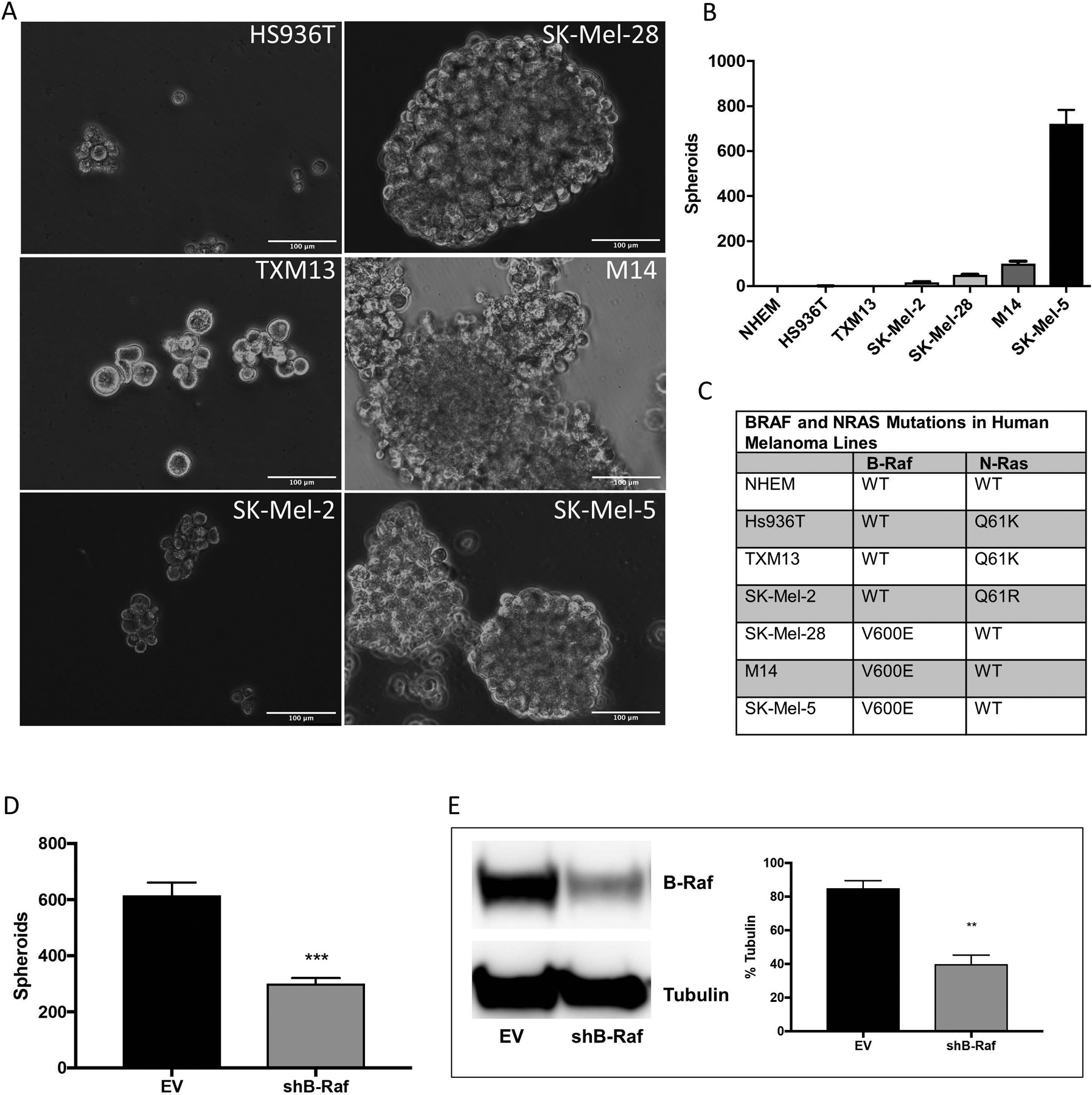
Figure 1. Melanoma cell lines harboring the B-RafV600E mutation form melanospheres.

Six human melanoma cell lines and primary normal human embryonic melanocytes (NHEM) were plated for melanosphere formation assays as described in Materials and Methods. After 10 days, the spheroids were imaged (A) and counted (B). (C) cDNA synthesized from RNA isolated from melanoma cell lines and NHEM was amplified by PCR and then N-Ras (NM_002524) and B-Raf (NM_004333) were sequenced. (D) SK-Mel-5 cells stably expressing either an empty pSicoR vector (EV) or a silencing hairpin RNA (shRNA) targeting B-Raf (shB-Raf) were plated for melanosphere assays. (E) B-Raf protein levels were assessed by immunoblot analysis using β-tubulin as a loading control. Error bars represent the standard error of the mean (SEM) of three biological replicates for immunoblot quantitation and four for melanosphere assays. Statistical significance was determined using the standard Student’s t-test. **P < 0.01, ***P < 0.001.
