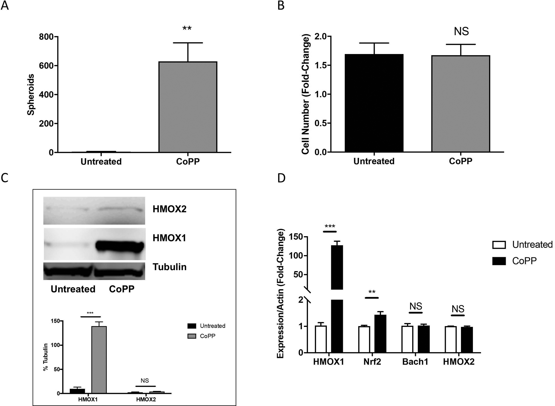
Figure 7. CoPP treatment facilitates melanosphere formation in Hs936T cells.

(A) Melanosphere formation was assessed in Hs936T cells treated with or without 10 μM CoPP. Error bars represent the SEM of four biological replicates. (B) Following the 10-day melanosphere assay, all cells were collected and melanospheres were dissociated by incubation with TrypLE and cells were counted. (C) Protein levels for HMOX1 and HMOX2 were assessed by immunoblot analysis for Hs936T cells grown in adherent conditions and either left untreated or treated with 10 μM CoPP overnight. Band intensities were measured and normalized to tubulin. Error bars represent the SEM of three biological replicates. (D) Expression levels of HMOX1, HMOX2, Nrf2, Bach1 and actin mRNA were assessed in Hs936T cells using qRT-PCR. Error bars represent the SEM of three biological replicates. Statistical significance was determined using the standard Student’s t-test. **P < 0.01, ***P < 0.001, NS is not significant.
