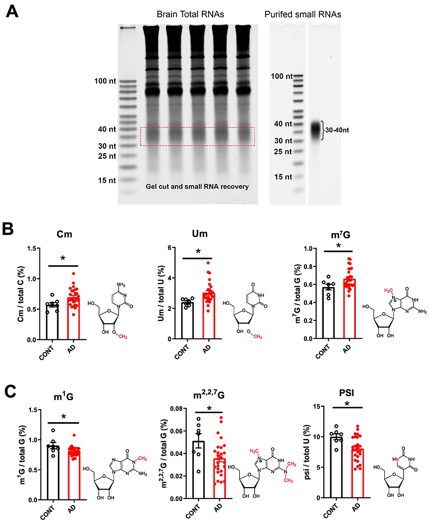
Figure 3.

Small RNA modifications in the 30–40-nt fraction from samples of the prefrontal lobe cortex of dementia patients. (A) Representative image showing purification of 30–40-nt small RNAs for the analysis of RNA modifications. (B) Increases in 2’-O-methylcytidine (Cm), 2’-O-methyluridine (Um) and 7-methylguanosine (m7G) modifications, and (C) reductions in 1-methylguanosine (m1G),N2,N2-dimethylguanosine (m2,2G) and pseudouridine (psi or Ψ) modifications in the cortex of AD compared with CONT subjects (each dot in the figure represents value from one independent sample); *P < 0·05 vs. CONT by Student t-test.
