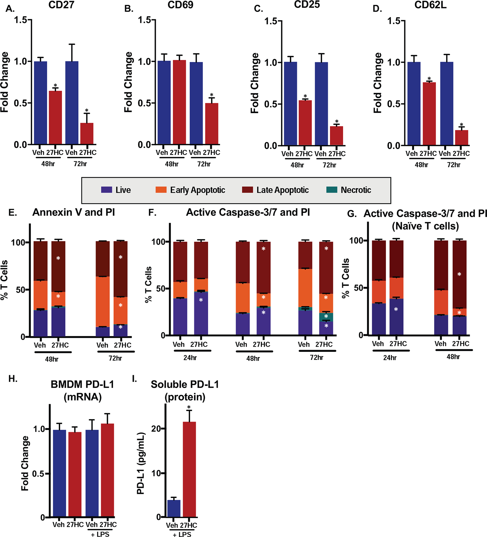
Figure 6: 27HC-treated macrophages induce T cell apoptosis.

(A-D) Gene expression of markers of T cell activation was downregulated when T cells are co-cultured with BMDMs treated with 27HC. qPCR on T cells, 48 or 72hrs after co-culture (N=3–4/group). Data are presented as normalized to respective vehicle control. (E) OT-I T cells 48 or 72 hours after co-culture with vehicle- or 27HC-treated BMDMs exhibited enhanced apoptosis. T cells were harvested from the co-culture at two different time points and then were labeled with Annexin V-FITC and PI. Flow cytometry analysis was performed to measure the cell populations (N=4/group). (F) OT-I T cells in co-culture with 27HC-treated BMDMs exhibit increased apoptosis. T cells were harvested from the co-culture (as in E) at three different time points and then labeled with FLICA-FAM, a caspase-3/7 inhibitor that covalently binds to active forms of caspase-3/7, and PI. Flow cytometry analysis was performed to measure the cell populations (N=4/group). (G) Naïve CD4+ T cells were isolated from the spleen, chemically activated and cultured with conditioned media from vehicle or 27HC treated BMDMs. Subsequent analysis was similar to (F) (N=4/group). (H) PD-L1 mRNA expression in BMDMs; see Fig. 1 for details. (I) Soluble PD-L1 is increased in media from LPS-primed BMDMs treated with 27HC for 24hrs (similar experimental setup to Fig. 4D (N=4/group). Data are presented as mean ± SEM, and asterisks (*) indicate statistical significance from the respective vehicle control. (two-tailed t-test in (A-D), two-way ANOVA followed by Bonferroni’s multiple comparison test in (E) and (F), p < 0.05)
