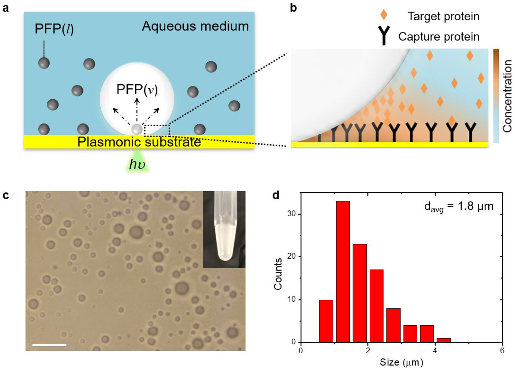
Figure 1. Scheme of bubble-enhanced surface capture of proteins and description of biphasic fluid.

(a) Schematic illustration of the bubble-generating PFP-in-water system and (B) bubble-mediated concentration of target proteins near the bubble/substrate interface. Arrows in (a) indicate the expansion of the PFP droplet into the bubble. (c) Optical image of PFP droplets (scale bar: 10 μm). Inset of (c) is a photograph of PFP-in-water fluid. (d) Size distribution of PFP droplets with a total number n=100.
