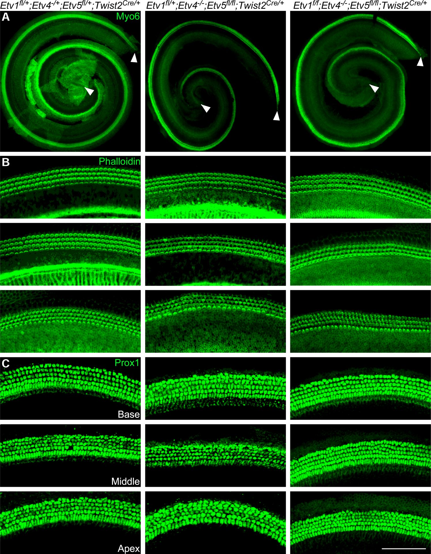
Fig 7.

Mesenchymal Etv1 is dispensable for cochlear development Whole mount immunostaining of P0 cochlea from control (Etv1fl/+; Etv4−/+; Etv5fl/+;Twist2Cre/+), Etv4/5 double mutant (Etv1fl/+;Etv4−/−;Etv5fl/fl;Twist2Cre/+) and Etv1/4/5 triple mutant (Etv1fl/fl;Etv4−/−;Etv5fl/fl;Twist2Cre/+) showing whole cochlea stained with Myosin 6 antibody (green) (A), and representative regions from each cochlear turn stained with F-actin (green) (B), and Prox1 staining (green) (C) as a marker for supporting cells. Scale bar=100μm. White arrowheads mark the beginning and the end of the cochlear length measurement.
