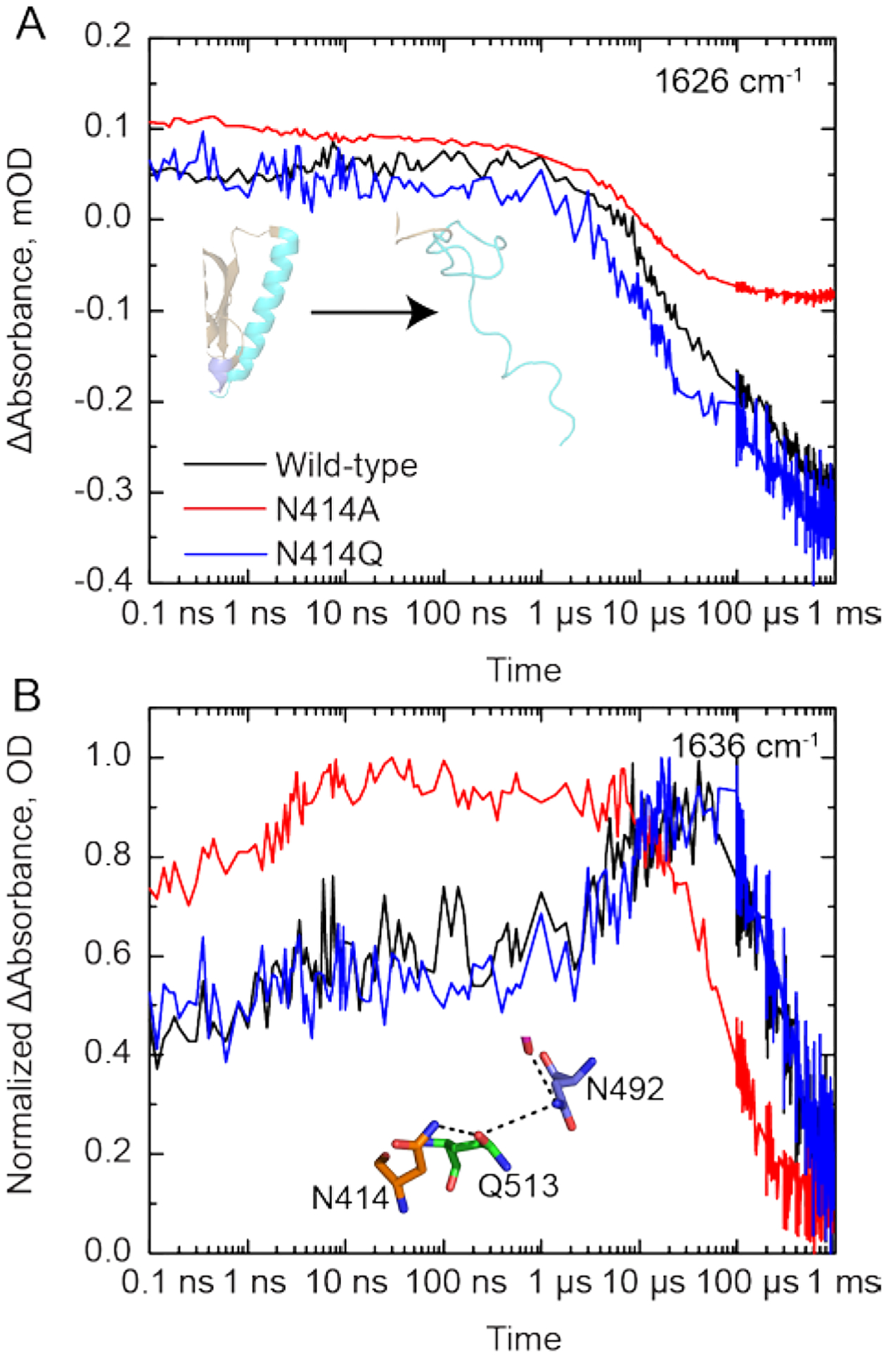
Figure 4: Selected kinetic traces from TRIR spectra of wild-type and mutant AsLOV2.

(A) The unfolding of the Jα helix is tracked by the increase in bleach intensity at 1626 cm−1 for the wild-type (black), N414A (red) and N414Q (blue) AsLOV2 proteins. (B) A rise and decay of the signal at 1636 cm−1 is assigned to structural dynamics associated with a transient hydrogen bond between N414 and Q513 due to the rotation of Q513. This signal decays to zero with the time constant of the fourth EAS.
