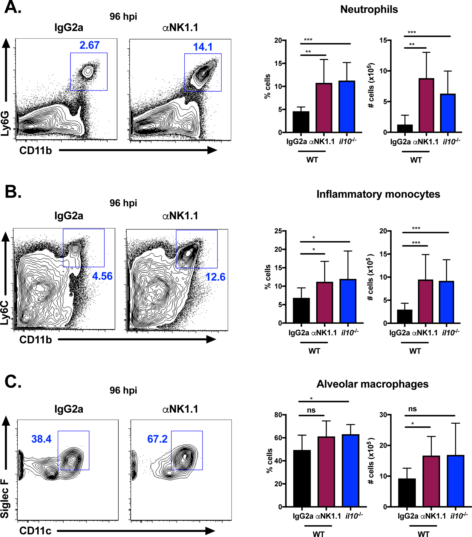
Figure 3. NK cells and IL-10 limit innate immune cell recruitment to the lung.

Representative flow plots of neutrophils (Ly6G+CD11b+) (A), inflammatory monocytes (Ly6G− Ly6ChiCD11b+) (B), and alveolar macrophages (Siglec F+CD11c+) (C) at 96 hpi in the lungs of WT mice treated with isotype control (IgG2a) antibody or αNK1.1 antibody 24 h prior to infection or il10−/− (untreated) mice infected with S. pneumoniae (107 CFU/mouse i.n.). Summary of cell percentages and total numbers for each group is also shown. Data are pooled from three independent experiments with n = 3–5 mice/group, *p<.05, **p<.01, ***p<.001 as measured by t-test.
